Videos
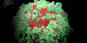
The Challenge of Creating a Vaccine for HIV
NOV 27, 2018 2:27 PM PST
Share
A Genetic Switch During Inflammation
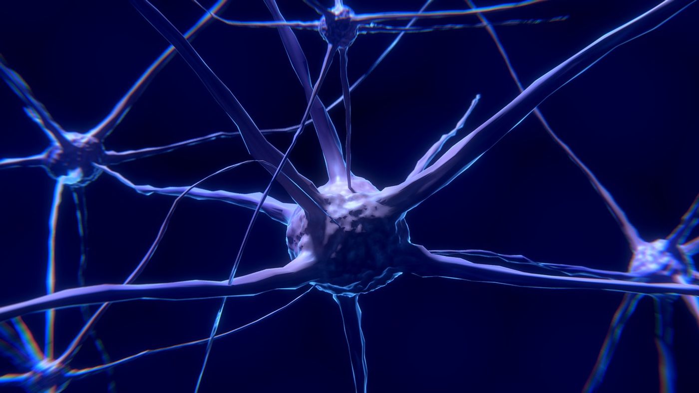
Wouldn’t it be great to better understand the mechanisms at play behind the scenes with regards to neurodegenerative diseases such as Alzheimer’s disease, Parkinson’s disease, multiple sclerosis, and brain cancer? Studies have been conducted to analyze these diseases; however, the activity of neuronal cells such as microglia during acute neuro-inflammatory processes such as those caused by infection, remain to be described.
A recent study performed at the NorLux Neuro-Oncology Laboratory at Luxembourg Institute of Health’s Department of Oncology reveals distinct gene expression profiles for microglial cells by way of single-cell-transcriptomics. Differences in genetic profiles are seen at steady state conditions, acute inflammatory conditions and neurodegenerative diseases. The details surrounding the observations with this study might accelerate future studies on therapeutic approaches to reestablish the central nervous system (CNS) cell function.
Microglia are specialized parenchymal‐resident phagocytes of the CNS that actively support, defend and modulate the neural environment. In other words, the microglia are surveying their surroundings to modulate the neural environment and protect against infection, toxin, or other contamination to promote brain health — Microglia coordinate immune responses between the periphery and the CNS as they perceive and propagate inflammatory signals initiated outside the CNS. Dysfunctional microglial reactions are thought to worsen CNS diseases.
The collaboration at the LIH between Dr. Alessandro Michelucci and Dr. Carole Sousa has facilitated the findings published in the November 2018 publishing of EMBO. Using single‐cell transcriptomics and multicolor flow cytometry this study presents comprehensive profiles of microglia in LPS‐injected mice, providing insight into microglia heterogeneity, and establishing a resource for the identification of specific phenotypes in CNS disorders. LPS stands for lipopolysaccharide. In this study, it is used to induce inflammation in the mouse model for observation. LPS works for this reason because it is what immunologists commonly refer to as a “PAMP” or pathogen-associated-molecular-pattern. Other common PAMPs include porins, peptidoglycan, lipoteichoic acids, mannose-rich glycans, flagellin, bacterial and viral genomes, mycolic acid, and lipoarabinomannan.
The researchers determine that microglia homeostatic signatures are mainly lost upon systemic inflammation, inflammation-induced microglia segregate into two distinct states and inflammation-induced microglia signatures are different from neurodegenerative disease-associated profiles. Disruption of CNS homeostasis, neuronal deterioration and inflammation are common pathophysiological features of several neurodegenerative diseases.
The researchers observed a marked global downregulation of the typical microglial homeostatic signature and simultaneously an up-regulation of genes classically activated by inflammation. 'When investigating further and comparing to published data, we could show that when being under acute systemic inflammation, microglia presented a highly activated state that is distinct from neurodegenerative disease-associated profiles,' states Dr. Sousa, who performed most of the experimental work.
Interestingly, the team observed heterogeneity among the activated cells, to which they hypothesize there may be a subset of reactive microglia less sensitive to the LPS stimulus. The findings will, hopefully, contribute to the establishment of new resources to unveil specific responses to brain disorders. Dr. Michelucci shares, “this should boost the development of novel therapeutic strategies against CNS diseases with an immunological component.”
For more information surrounding the basics of inflammation, watch this video.
Sources: EMBO, Science Daily, YouTube

Master's (MA/MS/Other)
You May Also Like
SEP 26, 2025
Cancer

Cancer immunotherapies, anti-cancer treatments that target a patient’s own immune system, working to make it more
...
NOV 26, 2025
Genetics & Genomics
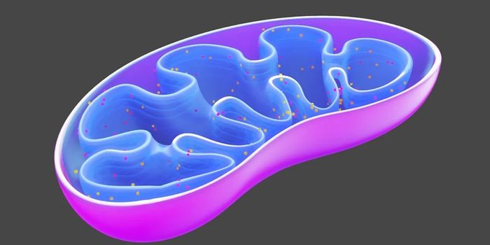
Mitochondria are well known as the powerhouses of the cell because of their energy generating capabilities. These little
...
DEC 11, 2025
Immunology

Scientists at the Van Andel Research Institute found that reducing caloric intake can help immune cells fight cancer. Co
...
DEC 18, 2025
Immunology

Immunotherapy has transformed healthcare and how physicians treat patients. Various forms of immunotherapy have been dev
...
JAN 01, 2026
Immunology
Immune cells work to fight infection and other diseases. Different subsets work together to elicit a healthy immune resp
...
JAN 15, 2026
Immunology

The concept of immunotherapy or boosting the body’s immune system to target tumors was first practically applied i
...
Loading Comments...