Systemic sclerosis (SSc) or scleroderma is a rare, chronic, autoimmune disorder that hardens connective tissue and muscle in the body. As a result, this can generate complications associated with skin, blood vessels, and internal organs. The production of excess collagen and fibrosis eventually leads to irreversible tissue and organ damage. Symptoms include Raynaud’s phenomenon (discoloration and numbness of fingers and toes), skin thickening, joint pain, stiffness, and gastrointestinal (GI) issues. Patients can also experience lung and kidney problems resulting in shortness of breath and high blood pressure. Interestingly, not all patients with SSc experience the same symptoms. Currently, there is no cure for SSc and treatment focuses on controlling symptoms. Regimens to mediate SSc include immune suppression medication, physical therapy, and surgery. Unfortunately, since the disease is so rare and has underlying complexities, scientists are still unsure what generates different symptom experience among patients.
A recent article in Nature Communications, by Dr. Masayuki Nishide and others, found a cell signature or pattern that helps explain the variation of symptoms in patients. Nishide is a physician-scientist and Director of Clinical Immunology at The Osaka Hospital and faculty member at the University of Osaka in Japan. His work focuses on specialized immune cells known as ‘granulocytes’ and how they function in disease.
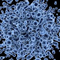
Nishide and his team discovered that the variation of SSc severity is due to the rapid proliferation and activation of specific immune cells throughout the body. In any autoimmune disorder, the dysregulated immune system is overactive, which causes vascular damage and tissue fibrosis. To investigate the variation of symptoms among patients, researchers took blood and tissue samples from SSc patients and analyzed the cells. The team documented gene expression and surface proteins to identify biomarkers for the disease.
Researchers discovered a specific immune cell type that express the gene EGR1. These cells are known as monocytes and are part of the innate immune system. In the cell analysis, these cells were strongly associated with scleroderma renal crisis – a serious kidney complication in patients with SSc. Normally, immune cells help fight infection, but the overactivation and targeting of ‘self’ tissues in this autoimmune disease generate deleterious outcomes for patients. The identified monocytes were found to transform into destructive cells that promoted chronic inflammation and generated thickening and scarring of internal organs. Additionally, the group found that another type of immune cell, known as T cells, had a gene pattern that altered their function to be more aggressive and inflammatory. In this case, these T cells were correlated with progressive interstitial lung disease. Researchers believe that the monocytes and T cells collectively contribute to the progression of SSc by not only increasing inflammation and causing tissue fibrosis, but also by recruiting other factors that drive the disease.
The discovery of different immune cell subtypes and their accumulation around the lungs and kidneys helped scientists explain symptom variation among patients. The quantity of these different cell types correlates to symptom severity. Additionally, these new findings provide insight into the underlying mechanism of SSc. Consequently, scientists can now move forward leveraging immune cell patterns to develop new therapeutic strategies and improve SSc treatment.
Article, Nature Communications, Masayuki Nishide, University of Osaka