The measles virus was caught leaving infected cells by the scientific paparazzi, that is, experts from Emory Health Sciences using cryo-electron tomography (cryo-ET). In their new study, researchers show how obtaining images of the measles virus at this stage of infection will help them understand better the pathogen’s structure and function.
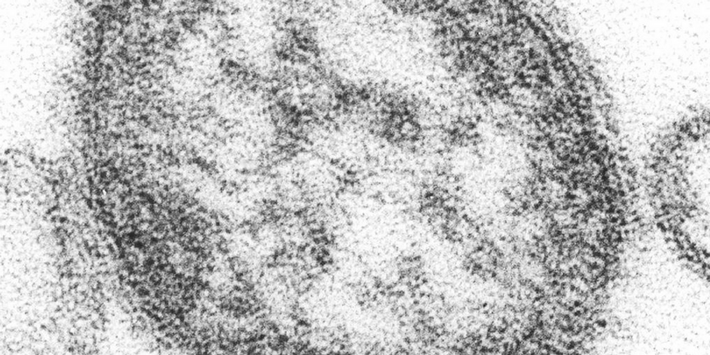
Similar to a CT scan, cryo-ET is an imaging technique that helped Emory scientists capture images of the measles virus, which is infamously difficult to work with as it is unstable and genetically diverse. It involves applying an electron microscope to take several 2D pictures of the virus while the sample is tilted at multiple angles along one axis. This rotation allows the technology to produce images that represent the virus’s 3D volume.
"With the whole-cell tomography approach, we can collect data on hundreds of viruses during stages of assembly and when released,” explained study co-leader Elizabeth Wright, PhD. “This allows us to capture the full spectrum of structures along the virus assembly pathway."
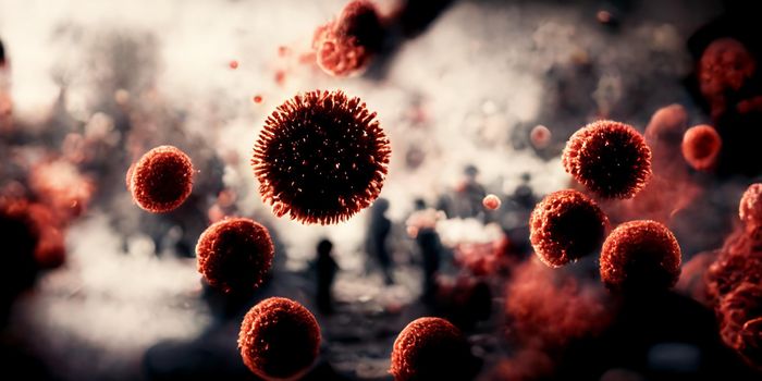
The measles virus infects the respiratory system, immune system, and skin, resulting in a very contagious infection that devastated the world before a vaccine was developed. Common symptoms include high fever, characteristic spots in the mouth called “Koplik” spots, malaise, loss of appetite, and a rash covering most of the body.
The images obtained from cryo-ET will help scientists map the internal organization of the measles virus, understanding more about its detailed functions, and how to target those functions to improve curative and preventative therapeutics. While there is a vaccine to prevent measles, scientists still don’t completely understand the virus responsible for the disease. The images will also help scientists in understanding similar viruses: parainfluenza, respiratory syncytial virus (RSV), and the Nipah virus.
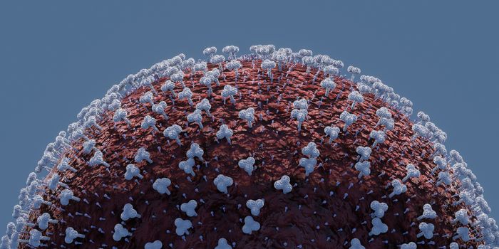
Via the cryo-ET images, researchers observed an internal matrix glycoprotein, the fusion (F) protein, forming a structural support system build on a foundation of interactions with the matrix (M) protein. They also saw visible “snakes” of encapsidated genetic material close to the viral membrane.
Additionally, researchers identified so-called “paracrystalline arrays” of M protein under the membrane that reportedly resemble Lego grid plates. These arrays have not been seen before in measles virus-infected cells or even the viral particles themselves.
While scientists will continue to glean important information from the images discusses in the present study, for now their findings prove that the structure of the measles virus may work differently than previously thought.
The present study was published in the journal Nature Communications.
Sources: Human Vaccines & Immunotherapeutics, Emory Health Sciences