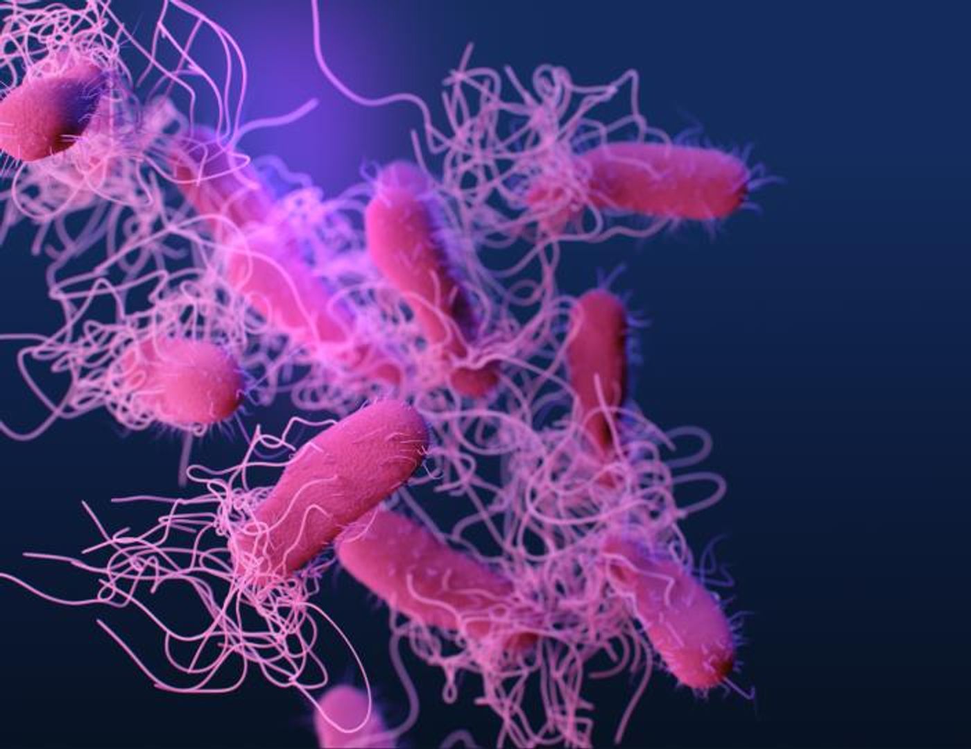
Researchers have created an imaging agent that will allow scientists and clinicians to visualize a bacterial infection caused by Enterobacterales pathogens while they're happening. These infections can affect many different parts of the body, are often resistant to treatments and may be life-threatening.
Enterobacterales are the largest group of bacterial pathogens that now impact humans, and they are known to sometimes cause dangerous infections that are hard to eliminate and can result in sepsis and death. This group includes Escherichia coli, Salmonella, and Klebsiella pneumoniae pathogens. They're also among the multi-drug resistant microbes that are considered a growing threat to public health.
It's been thought that if a non-invasive imaging tool can actually show clinicians where an infection is affecting the body, it would be easier to treat the infection. Now such a tool is being developed. The work has been reported in Science Translational Medicine.
Researchers have previously shown that when an easily synthesized substance called 18F-FDS (short for 2-deoxy-2-[18F]fluoro-d-sorbitol) is combined with an imaging tool called positron emission tomography/computerized tomography (PET/CT), the technique can be used to visualize Enterobacterales infections in a mouse model. Now, these scientists have shown that bacteria use a specific biochemical pathway involving the sugar sorbitol to absorb the 18F-FDS.
They also tested the technique on 26 volunteers who had various pathologies, some of which were confirmed Enterobacterales infections. The researchers showed that whole-body 18F-FDS PET/CT posed no risk, and it was able to rapidly illuminate where both drug-susceptible and multi-drug resistant strains of Enterobacterales were infecting the body. The tool also differentiated between these bacterial infections and cancer or inflammation caused by other things. The tool can show who is responsive to treatment and who is not; after patients with susceptible infections received antibiotic treatment, the signal intensity was reduced.
The work also showed that 18F-FDS PET/CT could tell the difference between SARS-CoV-2 and bacterial infection in a hamster model, so it may be specific to bacteria.
Experienced research scientist and technical expert with authorships on over 30 peer-reviewed publications, traveler to over 70 countries, published photographer and internationally-exhibited painter, volunteer trained in disaster-response, CPR and DV counseling.