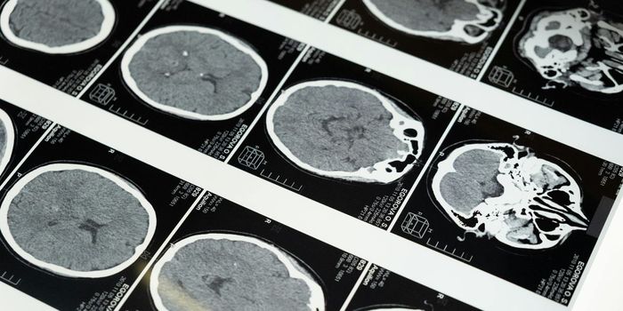
When neuroscientists and doctors have to diagnose complex brain illnesses or injuries, they must collect a great deal of information. From MRI scans to physical therapy evaluations, EEG studies and more, diagnosing conditions like Alzheimer's, autism, and Parkinson's disease can sometimes take months.
At the University of Rochester Medical Center (URMC) neurologists there have a powerful tool at their disposal that uses cutting-edge technology to look at these conditions in the brain and the rest of the body to hopefully come up with better ways to treat these devastating illnesses. The system, called the Mobile Brain/Body Imaging system, MoBI for short, uses motion capture technology, brain monitoring and state of the art imaging.
Dr. John Foxe, Ph.D., director of the URMC Del Monte Institute for Neuroscience, explained, "Many studies of brain activity occur in controlled environments where study subjects are sitting in a soundproof room staring at a computer screen. The MoBI system allows us to get people walking, using their senses, and solving the types of tasks you face every day, all the while measuring brain activity and tracking how the processes associated with cognition and movement interact."
The MoBI technology uses the same kind of tech that movie producers use to get stunning computer-generated images (CGI) that are used in blockbuster films. The team at URMC used it to study volunteers who donned black bodysuits fitted with a series of reflective disks. Once they are wired up, participants were asked to complete different tasks like walking, picking up objects, sitting, and standing in a room that had 16 high-speed cameras set up. The cameras can catch how and where the disks move within millimeters. The data points are then fed into a software program that develops a 3D map of all their movements. The investigators added environments like city neighborhoods for the testers to navigate and tasks that had to be completed based on pictures projected on the wall. EEG readings were taken via caps with electrodes on the scalp.
The information about what areas of the brain are active during the tasks was combined with the data points that showed movement and used to try and identify certain conditions. It's especially helpful in patients with autism. Many times someone with an autism spectrum disorder (ASD) will have difficulty processing stimuli from more than one source. Those with autism also often have an abnormal gait. In patients with Parkinson's or Alzheimer's muscle stiffness and eye-hand coordination are impacted along with cognition, so the MoBI system is able to track brain activity as well as mobility issues.
Dr. Ed Freedman, Ph.D., is an associate professor in the Del Monte Institute and principal investigator in the Cognitive Neurophysiology Lab where the MoBI system is housed stated, "There is competition between the processes that allow you to walk well and the processes that allow you to think well. We don't fall when we are sitting at a desk performing a task; we fall when we are walking down the street, avoiding traffic and other people, checking our phone, and thinking about what we are going to cook for dinner. We can use the MoBI to reveal underlying problems in the allocation of cognitive resources in individuals with neurological disorders because we are essentially stressing the system by asking them to perform a task while on the treadmill." Check out the video below for a look at the system.
Sources: University of Rochester Medical Center,