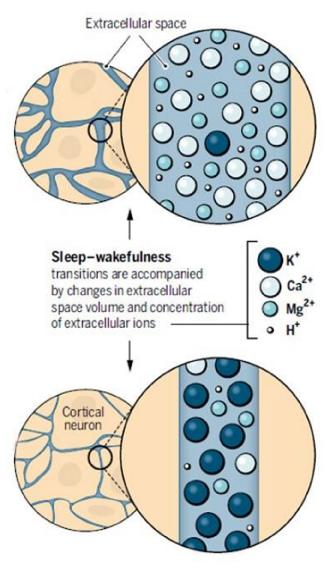
It is well-known that the extracellular concentrations of various ions in the brain change depending on the behavioral state, meaning awake or asleep. Sleep and wakefulness have their own distinct patterns of cortical electroencephalography (EEG) activity, gene expression,and metabolic activity. While awake, the level of extracellular K+ is much higher than when asleep. It was previously thought that this change in extracellular K+ was secondary to the increased neuronal activity during wakefulness, but new research suggests that the change in extracellular ions is a cause, not a consequence, of changing arousal states.
Researchers from the Center for Translational Neuromedicine at the University of Rochester Medical Center have recently published a paper in
Science that presents evidence for the changes in extracellular ions being the cause of the shift between sleep and wakefulness.
In addition to extracellular K
+, the researchers also looked at extracellular concentrations of Ca
2+ and Mg
2+. Ca
2+ and Mg
2+ are in the inverse of K
+, with higher levels during sleep that fall while awake. In the image above, the top part represents ion concentrations while asleep, and the bottom shows ion concentrations during wakefulness.
Most of the experiments in this paper were done
in vivo, in mice with a cranial window that allowed the researchers access to a small, local portion of the cortex. With the cranial window, they were able to apply artificial cerebrospinal fluid (ACSF) with a cocktail of neuromodulators that promote either sleep or wakefulness. When the wakefulness cocktail was applied to the cranial window of a sleeping mouse, it induced a rapid, local increase in extracellular K
+ and a slower decrease in extracellular Ca
2+ and Mg
2+ but did not wake the mouse. This effect was not dependent on local glutamatergic signaling, as determined by application of tetrodotoxin (TTX), an AMPA receptor blocker. The opposite was true as well, with the sleep cocktail being applied to the cranial window of an awake mouse locally lowering extracellular K
+ and raising Ca
2+ and Mg
2+.
In addition to these cranial window experiments, the researchers also tested if globally changing the ion concentrations would have a global effect on behavioral state. They infused sleeping or wakeful mice with ACSF with the cocktail to induce the opposite behavioral state. What they saw confirmed their cranial window experiments. They were able to wake up a sleeping mouse by infusing the wakefulness cocktail to globally raise K
+ levels and lower Ca
2+ and Mg
2+ levels. The opposite was true as well.
In addition to the changes in extracellular ion concentrations, the volume of interstitial fluid changes from awake to asleep. The volume and flow of extracellular fluid has been know to increase during sleep, a process that is thought to aid in clearing solutes from the brain. The researchers saw that changing the ion concentrations also changed the volume of extracellular fluid.
These results suggest that changing extracellular ion concentrations are a cause of behavioral state changes, not a consequence as previously thought. Since sleep disturbances have been implicated in numerous neuropsychiatric conditions, it is important to understand how our brains fall asleep and wake up. This knowledge will be crucial for developing therapies to better regulate these processes when they become disturbed.
Sources:
Science and
Alzforum