The expression “Seeing is believing” is one that most would agree with. The problem in neuroscience is that imaging in the brain, while useful, cannot capture everything. Brain injuries often need to be evaluated at the neuronal level and seeing these structures in the brain isn’t entirely possible…yet. Recent research from the Albert Einstein College of Medicine and the Montefiore Health System however made use of a new imaging technique that could predict concussion recovery rates as well as show how the brain goes about repairing itself.
Study leader Michael L. Lipton, M.D., Ph.D., professor of radiology, of psychiatry and behavioral sciences, and of neuroscience, as well as associate director of the Gruss Magnetic Resonance Research Center (MRRC) at Einstein and director of MRI services at Montefiore said in a press release, “Our study presents for the first time a precision approach to harness imaging at the time of concussion to forecast outcome a year later. While we still lack effective treatments, we now have a better understanding of the neurological mechanisms that underlie a favorable response to concussion, which opens a new window on how to look at therapies and to measure their effectiveness.”
According to the CDC, Traumatic Brain Injury (TBI) is a growing health concern In 2010, about 2.5 million emergency department (ED) visits, hospitalizations, or deaths were associated with TBI—either alone or in combination with other injuries—in the United States. Brain injuries contributed to the deaths of more than 50,000 people. It was diagnosed in more than 280,000 hospitalizations and over 2.2 million emergency room visits.
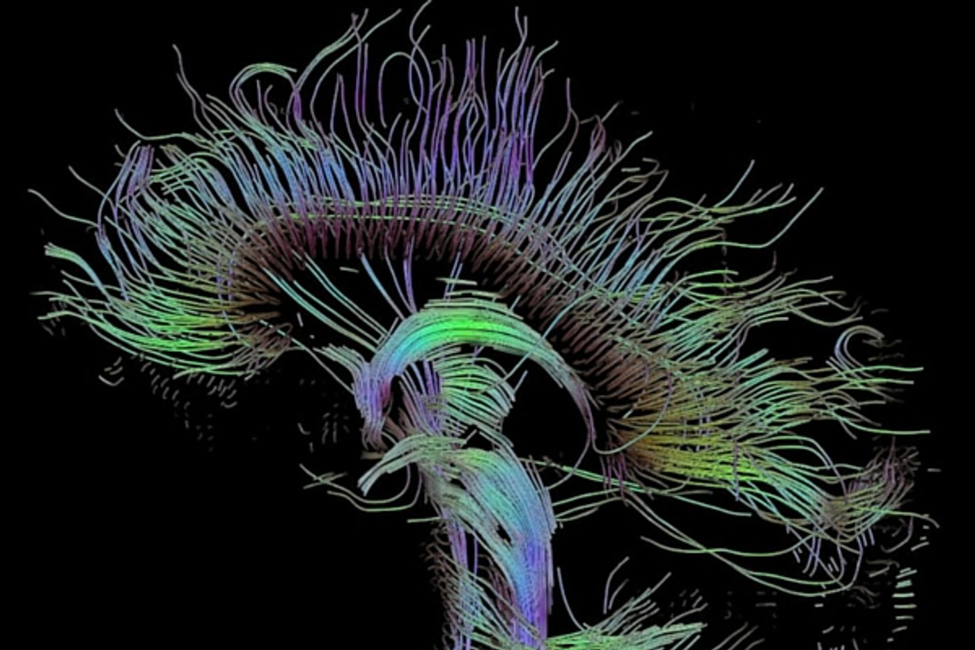
Concussion is all over the news in professional sports as well as in every day injuries. Diagnosing it can be problematic as it relies on evaluating symptoms, there is no hard and fast biomarker or objective assessment. MRIs and CT scans are often conducted, but they can't fully visualize damage done to the axons in the brain. These nerve fibers, that make up the brain’s white matter, are crucial to proper brain function but are vulnerable to concussion and other TBIs. The team at Albert Einstein used a newer form of MRI scans, diffusion tensor imaging (DTI), that can see the movement of water molecules in the brain. This movement, known as fractional anisotropy (FA) can indicate damage to the axons in an area of the brain. If FA is low, that indicates axonal damage.
“While most people think of concussions as a mild and short-lived injury, 15 to 30 percent of patients are left with symptoms that persist indefinitely,” said Sara Strauss, M.D., the study’s lead author and resident in the department of radiology at Montefiore. “Until now, we haven’t had a reliable way to differentiate in advance those who may be burdened long-term and those who would have a complete recovery.”
In the current study, Dr. Lipton compared the DTI scans of patients diagnosed with a mild TBI/concussion to those of healthy control subjects to see if they could offer reliable predictions about recovery. Patients were also assessed for three measures: cognitive function, post-concussion symptoms and health-related quality of life measures. A year later the concussion patients were re-scanned. The study found that the patients who had better outcomes a year later were also patients who had shown less areas of low volume FA in their initial scans. In addition, the patients who experienced more successful recoveries also had other brain areas with high levels of FA, indicating that the brain had adapted to the injury by forming fatty tissues around the injured area, allowing for fast nerve impulse movement.
Lipton stated, “Being able to predict which patients have a good or bad prognosis has tremendous implications for discovering and evaluating treatments for concussion. Developing an effective intervention requires first identifying the people who need it. Seventy to 85 percent of concussion patients get better by themselves, which makes it difficult to learn whether any treatment is actually helping. Our imaging technique allows researchers to test potential therapies on those concussion patients who can truly benefit from them.” Dr. Lipton explains in more detail in the video below, check it out.
Sources:
Albert Einstein School of Medicine,
CDC