Where does the ability to make a completely voluntary choice live in the brain? A recent paper published in Attention, Perception, & Psychophysics presents a novel experimental paradigm that claims to visualize the brain activity that accompanies a voluntary choice, or as they referred to it in the press release, free will. If the claim that scientists have visualized free will in the brain seems a little bold to you, you are right to be skeptical. That is not to say that this research isn’t novel or interesting, but such a strong assertion cannot be made with data from a single study.

This researchers from Johns Hopkins University used fMRI to look at brain activity while participants made voluntary decisions to switch their attention from one side of a split screen to another. Three major differences between this experiment and others that have tried to look at similar brain activity are 1) the participants had no instruction and were not responding to an external cue, 2) the switching of attention did not require any kind of movement, not even eye movement, and 3) the study did not rely on self-reporting. These differences are important because responding to an external cue or instruction or self-reporting a switch in attention by raising a hand activate their own neural networks, making it difficult to tease out which brain areas are involved in what processes.
By planning an experiment that eliminates external cues or self-reporting or movement, the researchers were able to get a clearer picture of what goes on in the brain leading up to and during a voluntary decision.
What they found was that the parietal cortex was involved in the actual attention-switching from one side of the screen to the other. This finding itself is not novel because many other studies have shown activity in the parietal cortex associated with attentional switching between spatial locations, features, objects, sensory modalities, task sets, and working memory representations. All of these studies had external cues, but this new study confirms that voluntary attention shifts and cue-evoked attention shifts are both processed in the parietal lobe.
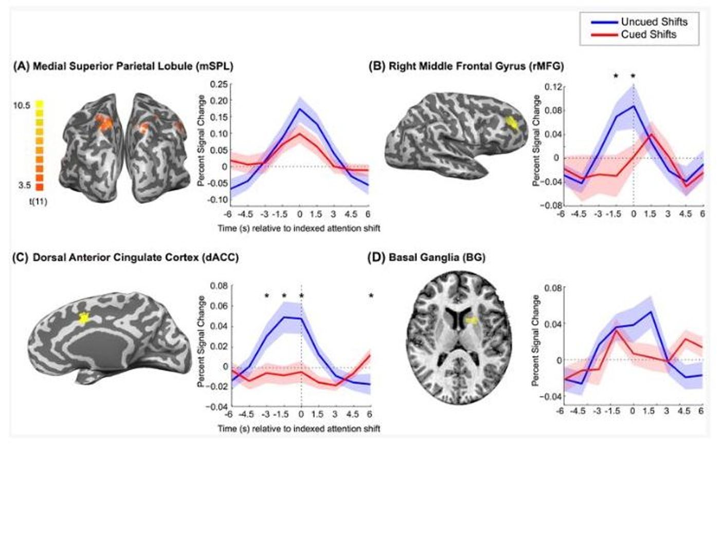
The more interesting finding was what the researchers saw in the lead-up to the voluntary decision. Compared to participants performing the same task but with an external cue telling them when to switch focus, the uncued attentional shift was preceded by an increase in activity in regions of the medial frontal and lateral prefrontal cortex, specifically in the dorsal anterior cingulate cortex and the right middle frontal gyrus. The researchers suggest that the activity in these areas “reflects processing related to the intention or preparation to reorient attention.”
This experiment is definitely cool and this protocol could potentially be used in the future to look at deficits in attention-switching that are associated with aging, substance abuse, ADHD, and a host of other neurological conditions. But the data has not convinced me that free will lives in the medial frontal and lateral prefrontal cortex. That is too strong of a claim. That said, this paper is exciting and will instigate more research into the roles that these brain areas play in choice and decision-making, and I look forward to following this line of research.
Sources:
EurekAlert and
AP&P