Epilepsy is the fourth most common neurological disorder in the United States. Patients who have it are of all ages and it can seriously limit one’s ability to enjoy life. It’s a spectrum disorder which means the kinds of seizures people suffer and how they are managed will vary depending on the patient. Currently about 3 million people in the US are living with epilepsy and experts predict that at least 1 in 26 people will develop epilepsy at some point in their lifetime. While epilepsy is most often treated with anti-seizure medication, there are some patients who have not benefitted from medication. This form of the disorder is called drug-resistant epilepsy and can be very difficult to treat.
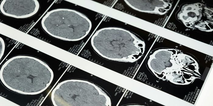
There are surgical options, but it’s crucial to have a good picture of the brain’s anatomy before any surgery is undertaken. Currently there are two methods for this. A recent article published by the American Academy of Neurology in the medical journal Neurology, looked at the two methods and revised the guidelines for each. The more common method of mapping the brain before surgery is the intracarotid amobarbital procedure, also known as the Wada test. In this procedure one side of the brain is anesthetized by injecting medication via the carotid artery. It’s invasive, can be uncomfortable and does carry some risk. The other way to get a look at the brain architecture is to use functional MRI scans.
Functional MRI is an imaging procedure that detects brain activity by measuring blood flow between regions of the brain. It’s considered much less risky than the Wada tests. Pre-surgical mapping of the brain is vital because the areas of the brain that process memory and language need to be known before operating, to avoid injury or damage. It is non-invasive and considered safe. The purpose of both tests is to ensure language and memory abilities will not be affected as a result of surgery.
Jerzy Szaflarski, MD, PhD, of the University of Alabama at Birmingham and Fellow of the American Academy of Neurology was the lead author of the article. He explained, “Because fMRI is becoming more widely available, we wanted to see how it compares to the Wada test. While the risks associated with the Wada test are rare, they can be serious, including stroke and injury to the carotid artery.” Still, there was only weak evidence to support the use of fMRI as a possible alternative for the Wada test for those patients with medial temporal lobe epilepsy, temporal epilepsy in general or extratemporal epilepsy. Additionally there was not enough good evidence to make a recommendation for people with temporal neocortical epilepsy or temporal tumors. For those patients considering left medial temporal lobe surgery there was some moderate evidence that fMRI scans could be used.
Epilepsy is a complex disease, and each patient must be evaluated based on their specific situation. The two procedures also differ depending on where an by whom they are performed. Having good information about the brain beforehand is crucial, so hopefully the new guidelines will help patients and doctors make the best decisions The guidelines were supported by the American Epilepsy Society and the American College of Radiology in addition to the AAN. The video below explains the two methods in more detail.
Sources: American Academy of Neurology, UPI, American Epilepsy Society