The lowly worm isn't usually considered an exciting or valuable animal. Many worms are stuck on the end of a hook for fish bait or added to a garden as fertilizers. However, one species of worm is now a rising star in the field of neuroscience.
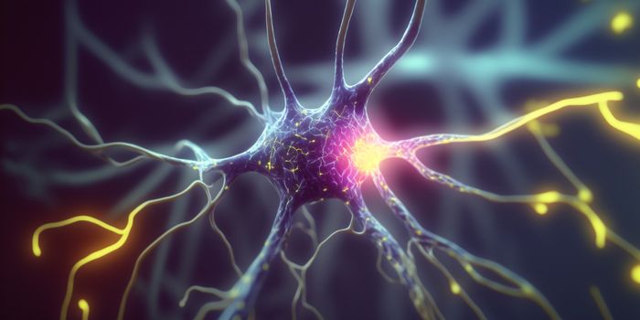
A crucial part of understanding how the brain works is seeing the neurons in action. In a human brain it's almost impossible to do at the single cell level. A human brain has over 100 billion neurons, and even the best imaging technology can't capture individual cells. When researchers want to get a closer look at how behavior might be related to neuron activity in the brain, the tiny worm species known as Caenorhabditis elegans or C. elegans is the new rock star. In research at Princeton in 2015 and more recent work from the University of California, San Francisco the neuron activity in these tiny transparent worms is being connected to behaviors like sensing danger and getting away, finding food and even looking for a mate.
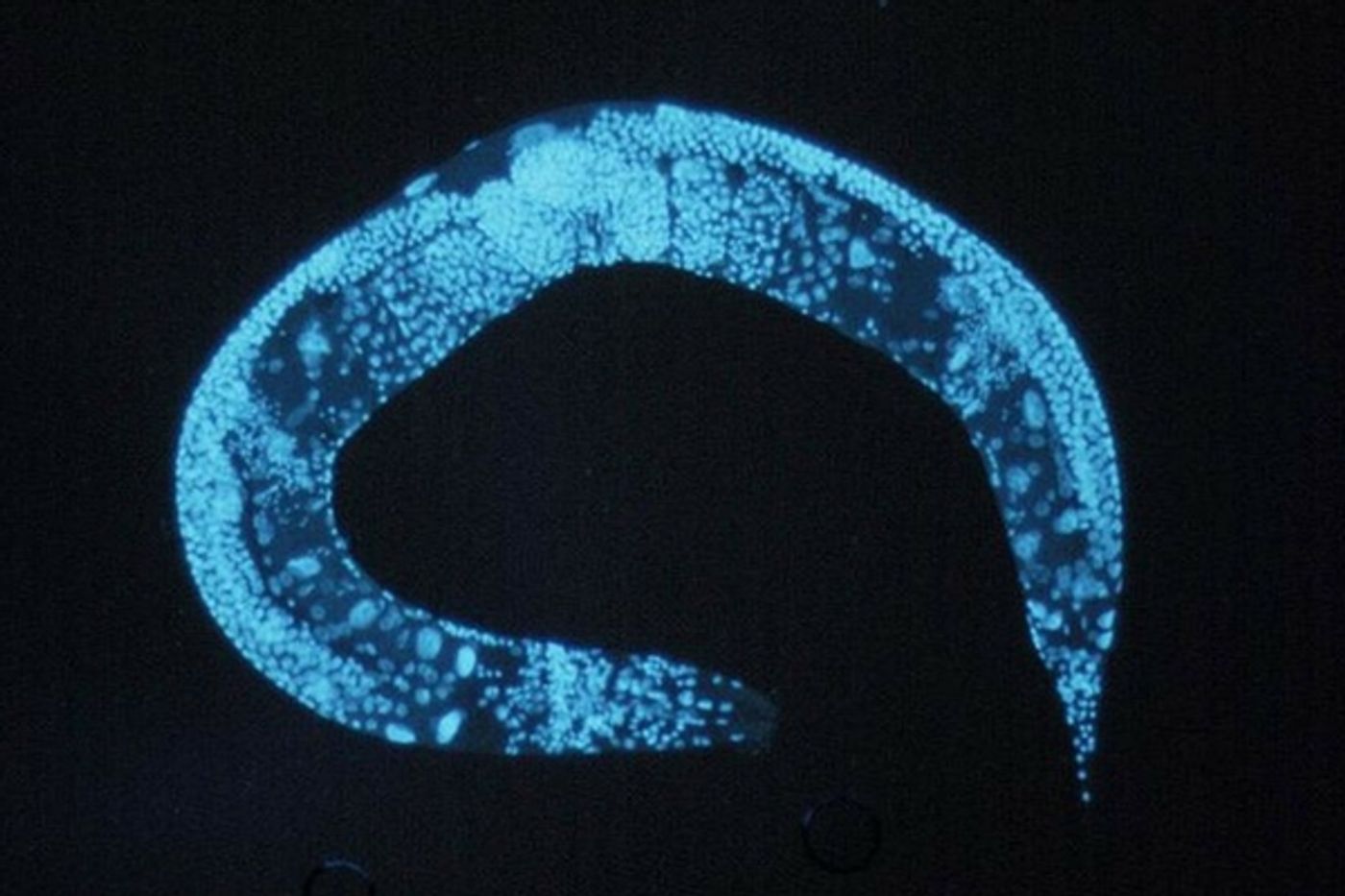
The most recent work is being led by Dr. Saul Kato, Ph.D., is an assistant professor of neurology at the UCSF Weill Institute for Neurosciences. In his lab, he's using a new imaging technique that allows him to see all 302 of the worm's neurons at the same time. C. elegans are less than one millimeter in size, and their brains are powered by a fraction of the number of neurons in most other animals. Kato explained, "They do everything all animals do, but they do it with only 302 neurons – and now we can watch nearly all of them at the same time."
Because neurons in other animals can't be seen all at once, some researchers would observe a particular group of neurons and then ascribe behaviors to that group's activity. The problem is that there was likely activity in other bunches of neurons at the same time, which contributed to behavior, but it went unseen. Kato's team at UCSF added a fluorescent calcium sensor to make the neuron's electrical activity visible under the microscope. When the concentration of calcium ions is changed, it corresponds to a change in the electrical sequences of neuron activity. Researchers were able to look at specific patterns and identify the behavior (food searching, turns and crawling) just by looking at the flashing of each neuron.
It was a very complex process to record the patterns of activity and match them to behaviors in the worms, so the next task for the team is to work on machine learning algorithms that can parse the electrical patterns quicker and more accurately. Dr. Kato compared the patterns of activity to a piece of music being sung by a chorus. To make sense of the song, it's necessary to hear all the parts. He stated, "This chorus was a surprising finding. We surmise that it is a signal telling each neuron what the body is trying to do so they can contribute meaningfully to the whole animal's function, like sailors on a submarine. It's a way for the neurons to communicate with each other."
The team at UCSF hopes to use the information they have gained by studying the C. elegans in finding out how disruptions in neural patterns are a factor in disease. Check out the video below to learn more.
Sources: UCSF, GlobalNewsConnect, Daily Mail