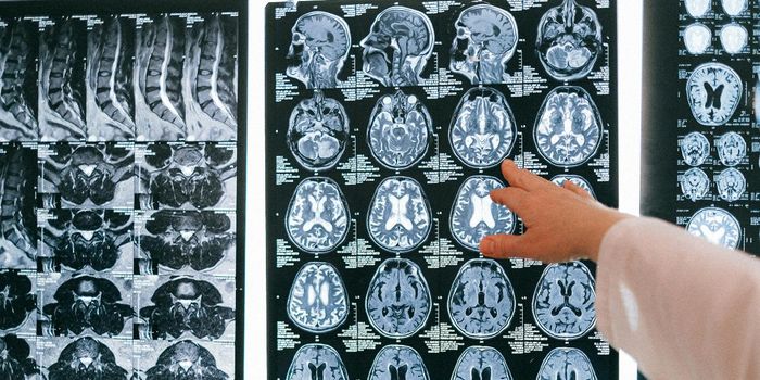
In the field of neuroscience, imaging is everything. Before the brain can be fully understood, it must be seen. The problem is that there are billions of neurons and imaging them is difficult. Even the most advanced techniques can only show a small amount of the brain at a time.
Functional MRI scanners (fMRI) are the new rock stars of the filed, but resolution remains a problem. The strongest MRI magnets are rated from 7 to 10 on the Tesla scale, but the best they can do is drill down to a region about the size of a grain of rice. A piece of the brain that small will still include about 100,000 individual neurons, engaged in a variety of different functions.
At the University of California, Berkeley, researchers there have come up with what they say is a way to up the ante on the current technology and increase resolution by a factor of 20. The school was recently awarded a $13.43 million grant from the BRAIN initiative at the National Institutes of Health to design and build what they call the "NexGen 7T."
David Feinberg, an adjunct professor at the Helen Wills Neuroscience Institute at UC Berkeley and president of Advanced MRI Technologies, the company who will lead the project, explained, "Our innovation in MRI technology requires a total redesign of nearly all of the scanner components, not just an incremental change. The much higher resolution imaging will overcome size barriers in imaging the cortex and should lead to new discoveries in the human brain, hopefully with major medical impact."
fMRI imaging, size matters very much. Rather than just revealing the structure of the brain and finding abnormalities in blood flow, size or position, fMRI scans show how the brain works and how each area is active in different ways. Patterns of neuron activity can be picked up by fMRI scans, but being able to see each part of the process is crucial. The new scanner should be able to image areas with a volume of 0.4 millimeters on each side and 2 millimeters long. This corresponds to the size of repeating microcircuits in the cerebral cortex. Each microcircuit is responsible for a different function in the brain, so being able to see each area separately will give researchers a better picture of the whole.
Size matters in the design of the scanner as well. While most traditional MRI scanners have a magnetic field, the size and variations of that field are essential. In the design of the NexGen 7T, the magnets are powerful, but rather than a few large coils of wire that pick up the signals, the new model's design calls for several smaller coils that can create a stronger signal and better resolution. This will allow researchers to look at layers of the cortex. The layers are an integral part of a scan since they reveal how the neurons are networked together, transmitting messages to other areas of the brain.
Professor Feinberg is a physicist, but the project will involve engineering experts, optometry specialists and computer scientists from Harvard University and Massachusetts General Hospital. The video below covers the basics of MRI technology which has been around since the 1940s but is continually being improved. Take a look at the science that goes into the fMRI technology.
Sources: University of California-Berkeley, Daily Californian, PsychCentral