While chronic traumatic encephalopathy (CTE) is a degenerative disease that builds over time, new research on high impact sports and young players shows that just one season of head-jarring play can cause changes to the brain.
Researchers at UT Southwestern Medical Center in Dallas and Wake Forest University recently presented the results of two studies that measured head impacts during one season and found that a specific region of the brain called the default mode network (DMN) was affected by playing the game, even when concussions didn't happen.
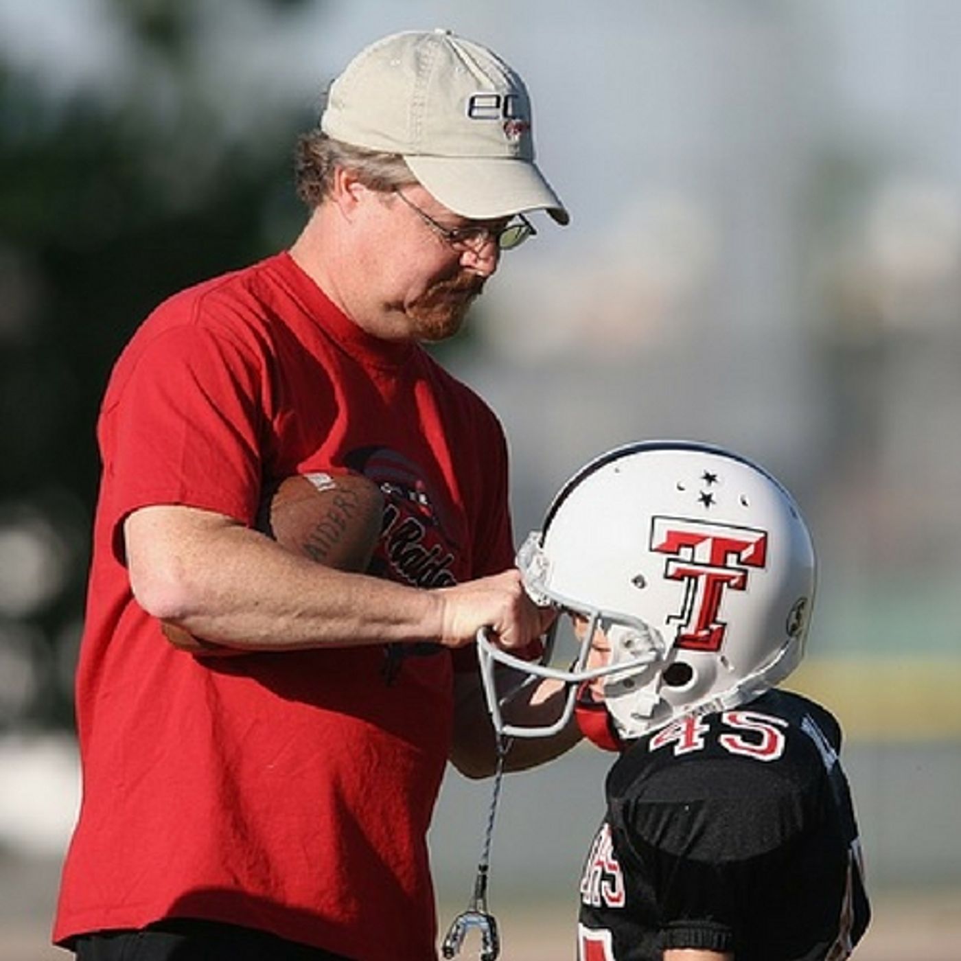
The DMN is a region of the brain that is engaged in times of wakeful rest. For instance, daydreaming or remembering an event and experiencing emotion in the memory. Patients with specific mental disorders show decreased connectivity in this network. Patients who have suffered a traumatic brain injury also have disrupted activity in this region. The first study involved 26 youth football players, ages 9-13. None had a previous history of concussion. Helmets for the players were equipped with the Head Impact Telemetry System (HITS). It's a series of electronic monitors and transmitters that record the metrics of any head impact.
The data collected during the season was analyzed to assess for risk of concussion. Players were divided into three groups: low exposure, high exposure and a control group of non-contact sports athletes.
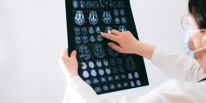
At the beginning and end of the season, each player underwent a functional MRI scan, and the connectivity of the DMN was explicitly noted. Gowtham Krishnan Murugesan, a Ph.D. student in biomedical engineering and member of UTs Advanced NeuroScience Imaging Research lab explained, "Over a season of football, players are exposed to numerous head impacts. The vast majority of these do not result in concussion. This work adds to a growing body of literature indicating that subconcussive head impacts can have an effect on the brain. This is a highly understudied area at the youth and high school level."
Using algorithms and machine learning, the data from the MRI and HIT system were analyzed, and the researchers could tell, with an 82% accuracy, which players were in the high exposure group. Murugesan concluded, "The brains of these youth and adolescent athletes are undergoing rapid maturation in this age range. This study demonstrates that playing a season of contact sports at the youth level can produce neuroimaging brain changes, particularly for the DMN."
The second study involved high school aged players, some of whom had suffered concussions. After wearing the HITS equipped helmets for the season these players underwent a magnetoencephalography (MEG) scan. These scans record the magnetic fields associated with electrical brain activity. When looking directly at the connectivity in the eight regions of the DMN, the results showed significantly decreased connectivity among the players who had suffered a previous concussion
UT postdoctoral researcher Elizabeth M. Davenport, Ph.D. stated, "The DMN exists in the deep gray matter areas of the brain. It includes structures that activate when we are awake and engaging in introspection or processing emotions, which are activities that are important for brain health. The brain's default mode network changes differently as a result of previous concussion. Previous concussion seems to prime the brain for additional changes. Concussion history may be affecting the brain's ability to compensate for subconcussive impacts." More information is in the video below, take a look.
Sources: Radiological Society of North America, UPI, CBS News