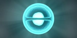
A recent developed microscope has allowed scientists to hold a front-row view on the “drama of mammalian development”. Scientists can now see inside a living mouse embryo, such as seeing the gut develop and heart cells take form. The study is "literally a cellular-resolution building plan of the entire mouse," explains a physicist and biologist, Philipp Keller, of the Howard Hughes Medical Institute's Janelia Research Campus in Ashburn, Virginia. Keller and research team published their findings in Cell. The researchers are making the microscope and computational tools along with all the imaging data to be free and publicly available.
The resources will be crucial for scientists seeking to grow or regenerate organs or even, someday, correct fix developmental problems that arise in the womb. "To do any of that, you first need to understand how organs form," she says. "You need to actually see what happens in a real embryo,” explains Janelia developmental biologist and study co-author, Kate McDole.
Until the present day research, viewing living embryos was only possible using certain species, particularly fish and flies. The viewing of embryos, since a decade ago, was through a “digital embryo” that Keller and colleagues once developed. However, using mice is completely different, because keeping mouse embryos alive is a challenge. The embryos need to be kept sterile and submerged in “nutrient soup” while gas and temperature levels are precisely controlled. Additionally, mouse embryonic cells are sensitive to light, their tissues are sometimes dense and opaque, and the embryo can't be still under the microscope, "drifts around like a little balloon," McDole explains. This is when the research team designed a “smarter” microscope with hopes to will address these challenges.

The smarter scope serves as one of the six scopes Keller and research team have developed and is a clear, acrylic cube housing the embryo imaging chamber. The scope has two light sheets illuminating the embryo and two cameras recording images. These characteristics of the smarter scope allow researchers to “spy” on the dynamic events in high resolution of the once-unseen world of early organ development. Additionally, the smarter microscope is equipped with a suite of algorithms that track the position and size of the embryo and can constantly adapt to change. "I wouldn't say our microscope is smarter than a human," Keller says, "but it's capable of doing things that a human operator would not be able to do. Without those programs, it would have taken a human two to three years to keep track of every cell.”
Source: Cell, Howard Hughes Medical Institute