In a study published in the journal Biomedical Optics Express, researchers have developed a capsule-shaped photoacoustic imaging endoscope that can allow physicians to have a better view of intestinal changes caused by Crohn's disease in affected patients—such information can advance treatment options.

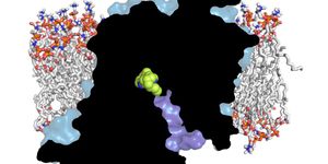
Caption: Researchers developed an endoscope that can perform photoacoustic imaging. The new device could give doctors a better view of intestinal changes caused by Crohn’s disease. Credit: Guan Xu, University of Michigan
"This new imaging technology could help more accurately plan therapy for each Crohn's disease patient," said Guan Xu, leader of the research team. "This would allow more targeted treatment and help minimize any adverse effects that might result from treatment."
The technology from the University of Michigan is used for photoacoustic imaging—a relatively novel biomedical imaging technique that uses light to produce sound waves in tissue which can be captured with ultrasound imaging.
Crohn disease leads to the development of strictures in the intestines which is caused by inflammation and fibrosis. Although the strictures that are a result of inflammation--can be treated with drugs, the strictures caused by fibrosis must be surgically removed.
"Currently, there is no imaging modality that can be used in the intestine to distinguish inflammation from fibrosis," said Xu. "The difficulty in accurately assessing the presence and development of fibrosis in the strictures adds a great deal of complexity to Crohn's disease management decisions."
The novel endoscope was tested on rabbit models and can examine whether the imaging technique could be used to characterize inflammation and fibrosis in intestinal strictures. It was designed to deliver infrared light at 1310 nanometers which allows to be absorbed by the collagen protein causing to expand lightly—a characteristic of feature of fibrosis.
"The method we demonstrated is minimally invasive and can directly assess fibrosis in the intestinal stricture, which has not been demonstrated by conventional medical imaging modalities," said Xu.
Source: Science Daily