A lens-free microscope devised by researchers at the University of California, Los Angeles (UCLA) detects cancer and other cellular-level irregularities with the precision of larger and more pricey optical microscopes, and is the first such instrument for high-throughput 3-D tissue imaging.
"This is a milestone in the work we've been doing," says Aydogan Ozcan, PhD, who serves as the associate director of UCLA's California NanoSystems Institute. "This is the first time tissue samples have been imaged in 3-D using a lens-free on-chip microscope."
The instrument uses a laser or light-emitting-diode to throw light on a tissue or blood sample on a slide. A sensor array on a microchip registers shadow patterns made by the sample and processes them as holograms, developing 3-D images of the specimen-which offers a virtual depth-of-field view. The reconstructed images are then color-coded by an algorithm, which sharpens the contrasts in the samples. The images that are hundreds of times larger in area compared with images captured by conventional bright-field optical microscopes, permitting faster specimen processing.
Ozcan's team tested the instrument using Pap smears that manifested cervical cancer, tissue specimens that included cancerous breast cells, and blood samples with sickle cell anemia. A board-certified pathologist analyzed specimen images in a blind test-some created by this lens-free technology, others by conventional microscopes. Diagnoses made with this new technology were unerring 99 percent of the time.
This microscope could pave the way for less costly and more transportable technology for viewing tissue, blood, and other specimens. "While mobile health care has expanded rapidly with the growth of consumer electronics-cellphones in particular-pathology is still, by and large, constrained to advanced clinical laboratory settings," Ozcan says. "Accompanied by advances in its graphical user interface, this platform could scale up for use in clinical, biomedical, scientific, educational, and citizen-science applications, among others."
Among Ozcan's lab's other innovations are bespoke smartphone attachments and apps that facilitate cell counts in blood samples, and using Google Glass to process diagnostic test results.
The research is the cover article in the most recent issue of Science Translational Medicine, published by the American Association for the Advancement of Science. The article, titled "Wide-field computational imaging of pathology slides using lens-free on-chip microscopy," can be found here: bit.ly/13m92Nl
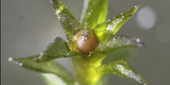
IMAGE: Tissue sample image created by a new lens-free microscope developed in the UCLA lab of Aydogan Ozcan. [Image courtesy Aydogan Ozcan, UCLA]