
Senior Manager, Global Field Applications Team, Ultivue
BIOGRAPHY
Innovative Biomarker Spatial Analysis for Comprehensive Tissue Immunophenotyping
Presented at:
Drug Discovery & Development Virtual Event Series 2023
Speaker
Abstract
Advances in tissue-based technologies over the past few years have created opportunities to identify patterns that can be used as biomarkers and prognostic indicators for different disease states. With the use of multiplex immunofluorescence (mIF) assays, the field has gone beyond visualizing a single protein or cell type on a tissue section and there is now room for greater complexity of image analysis. mIF can not only allow complex phenotyping of multiple cell types on a single tissue section, but the spatial relationships within the tissue can be both visualized and quantified. The incorporation of same slide H&Es, can even further increase the amount of information present on a single tissue section. This technology offers a unique advantage of preserving the architectural features of the tissue and revealing the spatial relationships that allow a detailed characterization of specific cell phenotypes defined by co- or lack of expression of multiple markers that may help in predicting clinical responses and mechanisms of resistance to treatment.
Learning Objectives:
1. Examine the concept that both spatial relationships and cell phenotypes can influence factors such as patient outcome and response to treatment.
2. Demonstrate how advanced image analysis software can be applied to discover cell types and populations within a morphological and spatial context.
3. Discuss real world examples of how whole slide image analysis can give insight into the immune environment and spatial relationships.
You May Also Like
FEB 17, 2026 | 8:00 AM

In cell therapy applications, commonly employed cell purification platforms for large-scale isolation of immune or stem cells separate cells based on a single cell surface antigen. Enrichmen...
FEB 18, 2026 | 8:00 AM

With advancing food allergy research and emerging therapies, new allergy testing requirements arise. In this context, Basophil Activation Testing (BAT) has emerged as an essential component...
FEB 18, 2026 | 1:00 PM

Energy-efficient filtration solutions that reduce hidden chemical exposure from benchtop work and lab equipment to improve overall laboratory safety...
FEB 19, 2026 | 7:00 AM
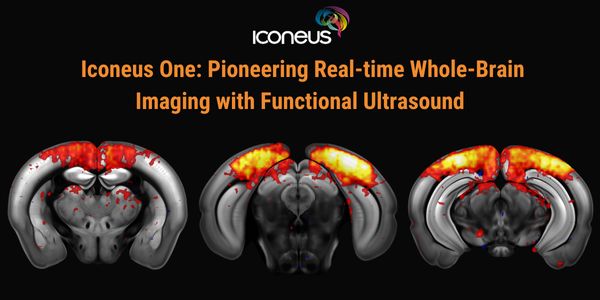
Discover how Iconeus One, the premier functional ultrasound (fUS) system for neuroscientists, is transforming brain function studies. Based on ultrafast plane-wave sonography, Iconeus One of...
FEB 19, 2026 | 8:00 AM

The collection of high-quality genomic DNA remains a major barrier in pediatric and neurodevelopmental research, particularly among children with autism spectrum disorder (ASD) and other neu...
FEB 23, 2026 | 9:00 AM
C.E. CREDITS

Explore decentralized liquid biopsy with SOPHiA GENETICS and MSK-ACCESS®, expanding access, improving outcomes, and accelerating precision oncology....
Loading Comments...