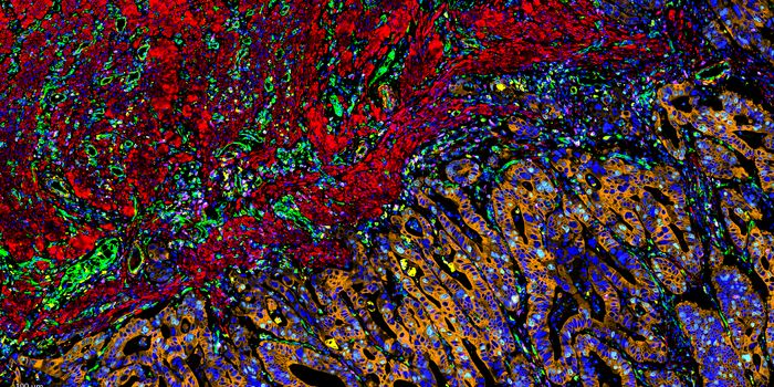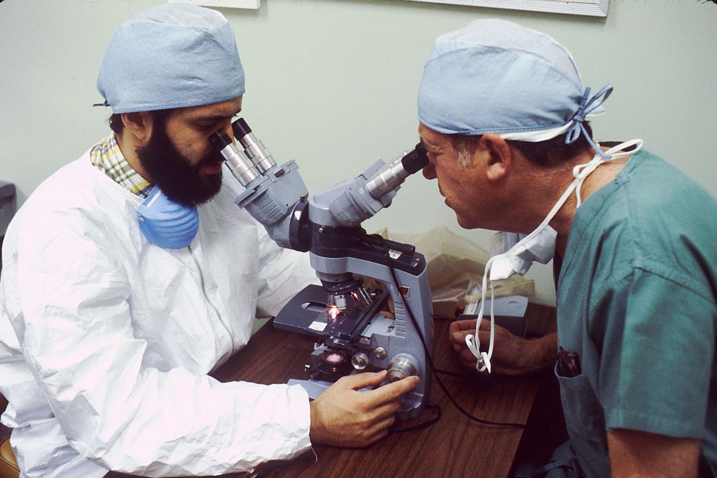
Histopathology describes the process of examining pieces of tissue using a microscope. Light microscopic (LM) examination of tissue helps diagnose several types of cancer by allowing pathologists to view cellular changes within a biopsy sample.
The workload of pathologists has increased in recent years due to policies that encourage screening for early cancer diagnoses. In addition, longer life expectancies and scientific advances have led to an increased number of cancer survivors, further increasing the need for pathology evaluations. Thus, strategies to efficiently utilize the limited pathology resources have become essential to maintaining standards of care and the health and safety of patients.
Digital pathology (DP) has emerged as an alternative method for analyzing tissue samples by stitching together digital images from histopathology slides. Automated slide scanners can rapidly generate these high-resolution images with minimal human interaction. In addition to the speed, DP does not require a microscope, offering remote viewing possibilities. Pathologists and other healthcare professionals can easily share images.
While small-scale comparisons of the accuracy of LM and DP suggest similar readouts, no definitive studies have demonstrated that DP can replace LM in the analysis and diagnosis of cancer.
A team of researchers throughout the United Kingdom sought to compare DP and LM in evaluating histopathology slides used to screen for various types of cancers. The researchers recently published the results of the comparison in Histopathology.
The study evaluated 2,024 cases (including 608 breast, 607 gastrointestinal [GI], 609 skin, and 200 renal cancers). A total of 16 pathologists participated in the study. Specifically, four pathologists specializing in the specific cancer type in question evaluated each case. Each pathologist viewed samples with both DP and LM with six weeks separating the examination methods. Clinical management concordance (CMC) indicated identical diagnoses.
For all cases (encompassing breast, GI, skin, and renal cancers), the CMC rate reached 99.95% when comparing LM versus DP. In addition, the study reported a 98.96% CMC rate for the samples used for breast and bowel cancer screening.
When evaluating site-specificity, all cancer types had high CMC rates for both overall and screening samples. Overall, CMC rates came out to 99.4%, 99.96%,99.99%, and 99.99% for breast, GI, skin, and renal cancers, respectively. The analysis of bowel and breast screening samples showed CMC rates reaching 99.93% and 96.27%, respectively.
The authors conclude LM and DP methods produce equivalent results when analyzing both routine and cancer screening samples.
Sources: Br J Healthcare Man, Histopath