Could stool samples reveal a person’s colorectal cancer risks? Researchers studying the molecular makeup of colon tissues and stool samples of mice say there’s a
metabolic fingerprint that’s distinctive for colon cancer. If the same results hold true for human stool samples, the scientists say a new non-invasive method to diagnose colorectal cancer could be on the horizon.

Colorectal cancer, also known as colon cancer or bowel cancer, is the third most commonly diagnosed cancer type in the world. The cancer usually begins as small, benign lumps of cells that form polyps in the colon. Without proper removal, these polyps can turn cancerous and cause symptoms such as abdomen pain, rectal bleeding, weakness, and fatigue. According to a recent report, the incidence of colorectal cancer is expected to increase by 60% to more than 2.2?million new cases and 1.1?million deaths by 2030.
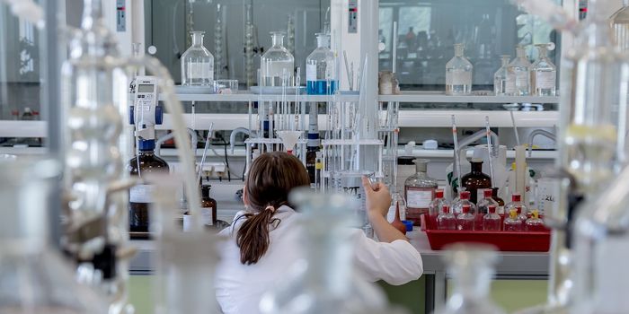
In attempting to find new ways to detect colon cancer earlier, researchers Washington State University and Johns Hopkins Medical School turned to a technology known as ion mobility-mass spectrometry (IMMS) and ultraperformance liquid chromatography. Essentially, this dual platform separates the molecular profiles of a biological sample, which can be interpreted as a metabolic fingerprint.
And as it turns out, these fingerprint are quite telling. By comparing the metabolic profile from normal colon tissues to those of cancerous polyps, the team found noticeable differences in many lipids and fatty acids. These markers created a molecular fingerprint for colorectal cancer in both human and mouse tissues.
The team next wondered whether fecal matter would also reflect these metabolic changes. Indeed, mouse fecal samples showed distinct metabolic abnormalities between those that were healthy versus those that were sick with colorectal cancer.
"The feces was not exactly the same as the tissue samples, but it had a lot of similarities to the tissue," said Herbert Hill, professor at Washington State University, and senior author of the study. "We found the lipids and fatty acids were changing -- and there were also changes in the amino acids."
Importantly, lysophospholipids were significantly altered in the colorectal cancer mouse model. "These types of lipids are known to be important in the development of cancer and are particularly tied to colorectal cancer,” said Hill
The evidence so far makes the team quite excited at the prospects of a noninvasive, early diagnostic test for colorectal cancer. "The exciting part is being able to see differences in the stool," said Hill. "This could lead to a noninvasive, more comprehensive early-warning detection method for colorectal cancer, but a lot of research needs to be done before it can be actually realized."
The team is gearing to test their method next in human fecal samples to see if the same fingerprints can be detected.
Additional sources: Washington State University via
Science Daily