How are new heart cells created? Is the adult heart capable of repairing itself after a heart attack? How can scientists intervene to improve recovery? University of California - Los Angeles (UCLA) scientists plan to answer all of these questions, but in their new Nature Communication study, they start with the first query.
A colorful new experiment used fluorescent proteins to study the development of heart muscle cells, called cardiomyocytes, as they grow in mouse embryos. The end game? Regenerate heart tissue in human adults to improve recovery after traumatic cardiac events like heart attack.
"Our ultimate goal is to be able to regenerate cardiomyocytes after an injury like a heart attack," explained Dr. Reza Ardehali. "But we're first trying to learn from the embryonic heart."
Ardehali and the other researchers chose to study cardiomyocytes because they are the main population of cells in the heart negatively impacted by a heart attacking, dying by the masses without an adequate supply of blood carrying oxygen and nutrients.
Cardiomyocytes are replaced, but not by other cardiomyocytes. Scar tissue takes its place, and the heart has to manage its regular duties but with less cells to get the job done. Studies show that in the adult heart, cardiomyocytes do not regenerate themselves; once they’re gone, they’re gone.
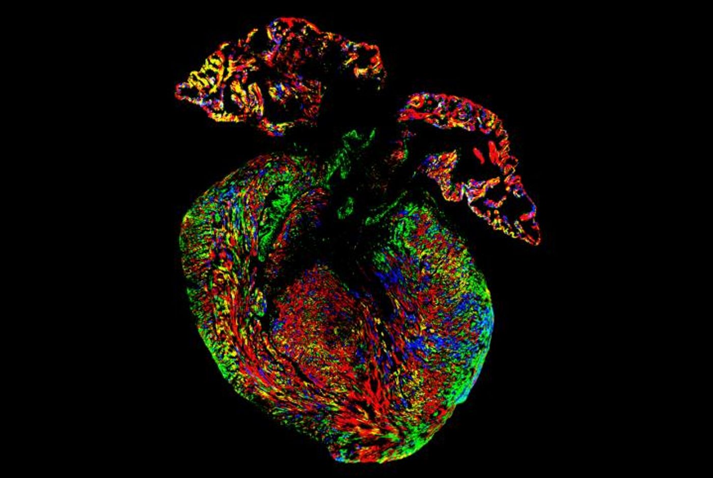
The research team from UCLA studied four different fluorescently-colored proteins to learn more about where cardiomyocytes came from in mouse embryos as they grew. Using fluorescent proteins meant that when cells divide, the daughter cells produced as a result maintain the same color. Researchers could easily keep track of the generations.
"Compared to previous labeling methods that use only one or two different colors, this method allows us distinguish the role of different cells much more clearly,” explained co-first author Ngoc Nguyen.
Cardiomyocytes and cardiac progenitor cells were among the types of cells labeled with fluorescent proteins. Progenitor cells are the “early descendents” of cells, capable of differentiating into different cell types. Researchers watched the colors change as they learned more about where cardiomyocytes were being produced during embryonic development.
They found that early in the development process, cardiomyocytes were more likely to originate from cardiac progenitor cells as opposed to other cardiomyocytes. However, cardiac progenitor cells gradually lose their ability to create new cardiomyocytes as development progresses. This is opposed to what scientists previously believed to be an abrupt transition from productive to unproductive progenitor cells.
"If we can determine how these cardiomyocytes proliferate, we hope to harness that regenerative potential for the treatment of heart disease in humans," Nguyen said.
Source: University of California - Los Angeles Health Sciences, Boston Children’s Hospital