Videos
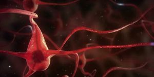
Mechanisms and secrets of Alzheimer's disease
APR 28, 2014 12:00 AM PDT
Share
Muscle Weakness Linked to Damaged Mitochondria
 People suffering from mitochondrial disease and those who have an extended history of alcoholism have the common symptom of muscle weakness. A study that was recently published in The Journal of Cell Biology appears to have unearthed the reason why.
Researchers from Thomas Jefferson University in Philadelphia discovered that the common thread is mitochondria that are incapable of repairing themselves. Mitochondria provide energy for virtually every cell in the human body, thus a reduction in functioning mitochondria could certainly explain muscle weakness.
Mitochondria typically repair themselves with a fusion process - combining with nearby mitochondria and blending components. The damaged components are swapped out with healthy protein sections from normal mitochondria, removed from the repaired structure, and isolated for future use.
However, skeletal muscles are tightly packed with fibers, with mitochondria compressed within the muscle structure. It has been assumed that the repair mechanism would be different within muscle, because of the lack of space and mobility to allow fusion to take place.
The research team suspected that fusion might still be a valid mechanism within muscle mitochondria, bolstered by the recent findings on two different mitochondrial disorders. The diseases ADOA (Autosomal Dominant Optical Atrophy) and a form of CMT (Charcot-Marie-Tooth) share muscle weakness as a symptom, and patients of these diseases have a mutation in a gene affecting the fusion of mitochondria.
For testing purposes, a strain of rats was created with mitochondria that always expressed a red color. This mitochondrial variation was also designed to change to a green color upon contact with a specific wavelength of laser output, thus producing a green square of mitochondria amid the background.
The team did produce the green squares as expected with laser activation, showing the exchange of contents with nearby red mitochondria. However, the "green" mitochondria were detected migrating within a muscle structure for significant distances, indicating fusion could take place there - and fusion did indeed happen.
Once the migration process and corresponding fusion was proven to be possible in muscle cells, could it be correlated to the muscle weakness mechanisms? The next step was to determine which of the proteins played a dominant role in the fusion inside skeletal muscle cells. With further research, the team determined that a protein labeled Mfn1 was the primary protein for the fusion process.
With the target protein identified, the team then tested the effect of alcohol. Rats that were given alcohol on a regular basis suffered from lower levels of Mfn1 - as low as half the amount in some cases. Mitochondrial fusion in these rats dropped drastically as a result. Replenishing the Mfn1 supply restored the mitochondrial fusion process, and reduced the muscle fatigue exhibited by the rats.
With the ability to turn the syndrome off and on via Mfn1 levels, the team proved the link between muscle weakness and mitochondria that could not repair themselves. In practical terms, since the dominant protein was identified, this work may lead to effective drug treatments against these forms of muscle weakness.

Bachelor's (BA/BS/Other)
You May Also Like
NOV 04, 2025
Cell & Molecular Biology

In recent years, effective obesity treatments like Ozempic and Mounjaro have changed lives. These drugs mimic the effect
...
NOV 09, 2025
Microbiology

It's been estimated that about one-third of all food that is produced for human consumption is wasted.
NOV 11, 2025
Cell & Molecular Biology

Astroviruses may not be well known, but they are very common and are the leading cause of diarrhea in children...
NOV 16, 2025
Cell & Molecular Biology

After an illness like the flu, symptoms may go away while a person still feels lousy. Researchers wanted to learn more a
...
DEC 16, 2025
Genetics & Genomics

Sometimes, a small error in the sequence of DNA can lead to a very serious disease. Scientists have identified many muta
...
JAN 13, 2026
Microbiology

Some recent studies have suggested that there is a link between severe gum disease or periodontitis, and some disorders
...
Loading Comments...