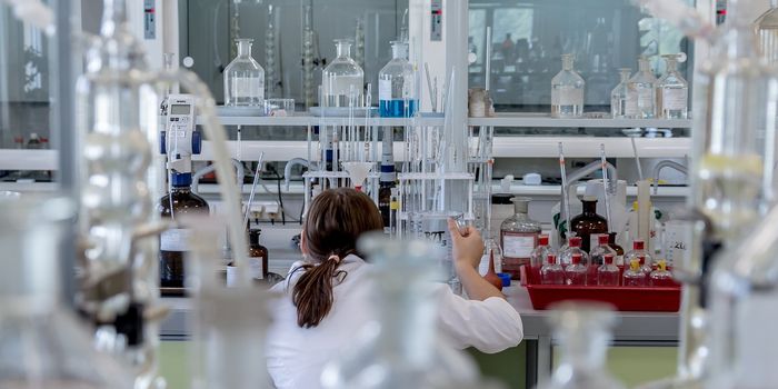
As the tools that scientists use to see cells and the structures inside of them have improved, our understanding of biological function has advanced. There are still limitations, however, and engineers are continuing to work to make microscopy techniques even better. Often, to see what’s happening in a cell, it must be fixed in place and studied after it has been preserved, but is no longer alive. Now researchers have created a method to see tiny things inside of live cells so that the dynamic events within can be viewed as they happen.
This technique, reported in Nature Communications, uses a chemical tracer that is already in wide use - heavy water or D2O. It's been combined with stimulated Raman scattering (SRS), a laser-imaging tool that was developed relatively recently. It may not only be useful in the lab; it could also help surgeons remove tiny bits of tumors.
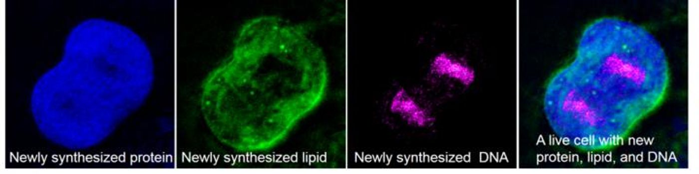
�"We can use this technology to visualize metabolic activities in a wide range of animals," said Wei Min, a chemistry professor at Columbia University and senior author of this study. "By tracking where and when new proteins, lipids, and DNA molecules are made, we can learn more about how animals develop and age, and what goes wrong in the case of injury and disease."
The water molecule has two hydrogen atoms and an oxygen atom; heavy water is made by using deuterium in place of the hydrogen. Anything under five tablespoons is not dangerous to humans, and cells will incorporate it into proteins, lipids and DNA they’re making.
Deuterium and carbon form a bond that vibrates at different frequencies when hit with light so that the various cellular molecules can be identified. The frequency signature a molecule emits allows it to be tracked through the cell. This labeling method is already used for molecules that are extracted, but this puts it into live cells.
"We get a continuous picture of what's happening inside living animal cells. Previously, we had only a snapshot," said the co-lead author of the work, Lingyan Shi, a postdoctoral researcher at Columbia.
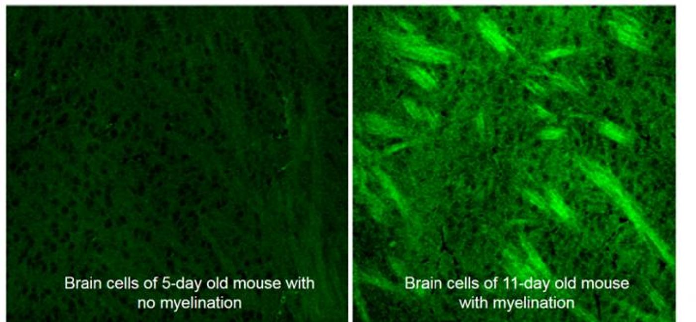
With this tool, the investigators watched the heavy metal get incorporation into parts of zebrafish, mice, and roundworms. They tracked fat production in the reproductive system of the roundworm as it aged. They observed the formation of a critical neuronal insulator called the myelin sheath in developing mouse brains, and other developmental events.
"The beauty of this mapping method is its simplicity," added Eric Potma, a chemistry professor at the University of California at Irvine who was not involved in the study. "It produces vivid images of metabolic activity in tissues with seemingly minimal effort. As the SRS microscope continues to get smaller, deuterium-labeled SRS imaging may help to catch tumors at much earlier stages."
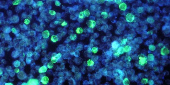
Sweat glands cell turnover gradually pushes older cells are to the center of the glands, expelling them from the body. Here, cells at the outer edges of the mouse sweat glands (upper left and lower right) appear bright green, reflecting new lipid growth. Older cells in an interior sweat gland (lower left) have far less new lipid growth. (Wei Min lab/Columbia University)
Sources: AAAS/Eurekalert! Via Columbia University, Nature Communications