By actually viewing live immune cells in action, scientists have learned more about they act as sentinels, keeping watch for pathogens. The team, led by Dr. Adam Wall and Dr. Nicholas Condon from Professor Jenny Stow's lab at UQ's Institute for Molecular Bioscience discovered a new structure on these cells that they called ‘tent-pole ruffles.’ These structures also shed light on how the growth of aggressive cancers is sustained.
"It's really exciting to be able to see cell behavior at unprecedented levels of resolution," Wall said. "This is discovery science at the cutting edge of microscopy and reveals how much we still have to learn about how cells function."
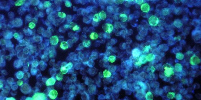
Reporting in the Journal of Cell Biology, the investigators used a tool called lattice light sheet microscopy. In light sheet microscopy, a laser beam is shaped into the form of a thin sheet, which passes through a specimen. Images are then detected in a different axis, which maximizes efficiency and minimizes background and damage to the sample. The lattice light sheet technique speeds up detection time to reduce damage even more, making it a great way to image biological processes as they happen. Learn more about it from the video.
The team studied a cell called a macrophage, which is a white blood cell that can find and gobble up any stuff that poses a potential threat to our health - that might be a microbe, a foreign substance, or a cancer cell. Macrophages can take up a lot of fluid, which helps them find unhealthy material, and can also trigger an immune response when necessary.
Their microscopy work enabled the researchers to discover the tent-pole ruffles on macrophages, which seem to help the cells sample the fluid around it. That sampling is called macropinocytosis. It was captured with unprecedented precision in only a few seconds.
The tent-pole ruffles are projections from the macrophage with a membrane between them as seen in the video; they allow the cells to capture especially big gulps of fluid, Wall noted.
This incredibly detailed imagery provides us with a new understanding of macropinocytosis and immune function, said Stow.
"This imaging will give us phenomenal power to reveal how cell behavior is affected in disease, to test the effects of drugs on cells, and to give us insights that will be important for devising new treatments" she added.
In cancer, for instance, the process of macropinocytosis is used by aggressive cancer cells as well. It helps them grab all the nutrients these hungry cells need for their rapid growth. The tent-pole ruffle structure is also seen in cancerous cells; the researchers plan to study molecules they have found on the ruffles, to see if they can use the ruffles to help impede cancer growth.
Sources: University of Queensland, Journal of Cell Biology