In research, it’s often critical to be able to see structures at the subcellular level. Scientists have been making improvements to microscopy techniques since the tool was invented, and a critical part of the process is preparing samples so they can be visualized with a microscope. Researchers at the University of Geneva have now created a method for seeing the structure of subcellular compartments called organelles; biological samples can be enlarged without altering them, and stuff that was once incredibly small can be seen in fine detail. The tool is powerful enough to allow researchers to see biochemical changes on protein complexes. It has been reported in Nature Methods.
Several years ago, a professor at the Massachusetts Institute of Technology (MIT), Edouard Boyden, had the idea to apply sodium acrylate, a common competent of baby diapers, to his research. ”Three years ago, Edouard Boyden developed a method to embed cell structures with a mixture of sodium acrylate and acrylamide, a chemical substance that is formed during the browning of French fries. He then marked the targets to be observed with fluorescent molecules before swelling the whole biological sample with water. The targets had to be destroyed, but it was possible to visualize their fluorescent borders with a good resolution, thanks to the expansion obtained,” explained Paul Guichard, Professor at the Department of Cell Biology of the UNIGE Faculty of Science.
This tool can now be applied to study the form of organelles, and how they function. The researchers used it to study organelles that act as a cellular power plant - mitochondria, and whether a part of the cell architecture, the centrosome, is part of them. "We investigated whether it was possible to adapt this technique to observe organelles without having to destroy them, and enlarge them without deforming them," said Virginie Hamel, a researcher at the Department of Cell Biology and co-author of the study.
After teaming up with colleagues at the University of Würzburg in Germany, the method was refined until they learned how to expand a sample while maintaining the structure, all without a chemical fixative that can change it. "Cells gradually expand, and their components separate from each other while enlarging. The architecture of the various elements is preserved and it becomes possible to observe them with a resolution hitherto unattained in optical microscopy,” noted the first author of the study Davide Gambarotto, a researcher in the Geneva group.
With this technique, called Ultrastructure Expansion Microscopy (U-ExM), protein complexes get anchored in a polymer. The polymer expands in a liquid, destroying protein interactions.
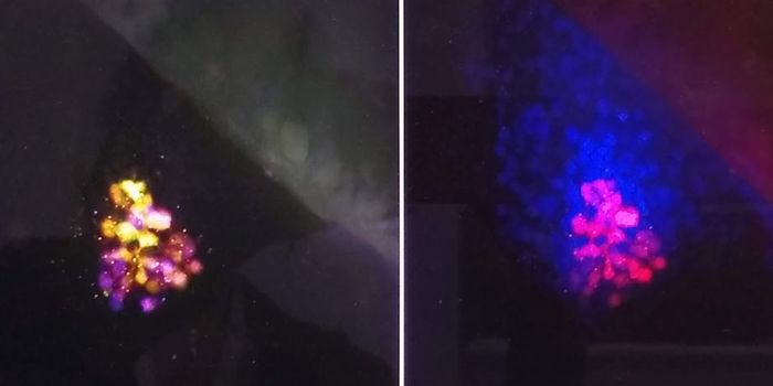
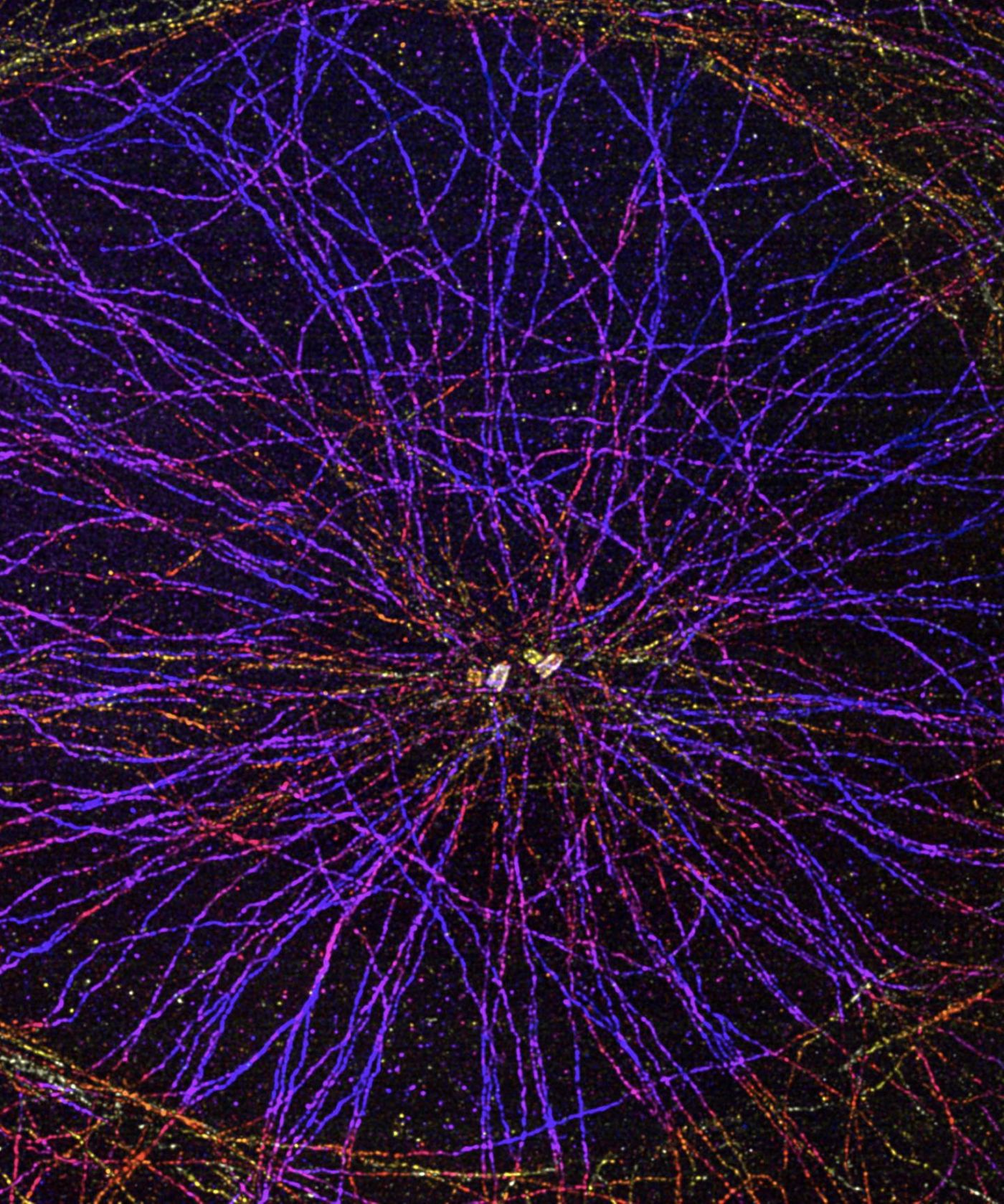
"The polymer then expands uniformly in all spatial directions by a factor of four. The antigens are retained and can subsequently be stained with dye-labeled antibodies," said Professor Markus Sauer of the Biocenter of Julius-Maximilians-Universität Würzburg. "With U-ExM, we can really depict ultrastructural details, the method is reliable, and it delivers a picture that is four times higher resolved than with standard methods of microscopy."
U-ExM can reveal details previously visible only through electron microscopy. “However, electron microscopy does not allow to locate the proteins that constitute the observed elements. Our method combines the advantage of fluorescence microscopy to detect molecules and high resolution to visualize the fine structure of organelles or of macromolecules,” noted Hamel.
"It is now becoming possible to map large intracellular molecular complexes. This method could also be used to reveal signatures of pathological processes at the very heart of the cell,” concluded Guichard.
Learn more about expansion microscopy from the video above, by MIT about work in the Boyden lab.
Sources: AAAS/Eurekalert! Via University of Geneva, AAAS/Eurekalert! via University of Würzburg, Nature Methods