Scientists have developed a tool for using gene expression to get an image of a cell, called DNA microscopy. The technique requires some simple methods as well as genetic sequencing and an algorithm. The genetic material in the cell is labelled, providing information about the spatial arrangement of cellular components. The researchers have used DNA microscopy to study human cancer cells, and they want to apply it to a variety of other cells in which small changes in genetic sequences have significant impacts, like immune cells.
"It's an entirely new category of microscopy," said Howard Hughes Medical Institute (HHMI) Investigator Aviv Regev. "It's not just a new technique, it's a way of doing things that we haven't ever considered doing before."
In DNA microscopy, short genetic sequences called unique molecular identifiers (UMIs) are tagged. A sample under analysis would be decorated with UMIs. Many of those tags will eventually collide with one another, and they will link together. Molecules that are closer together will join more frequently, and the ones that are farther apart won’t create as many connections. All of the interactions between the tags have to be genetically sequenced, and then analyzed with an algorithm, that assess about 50 million base pairs.
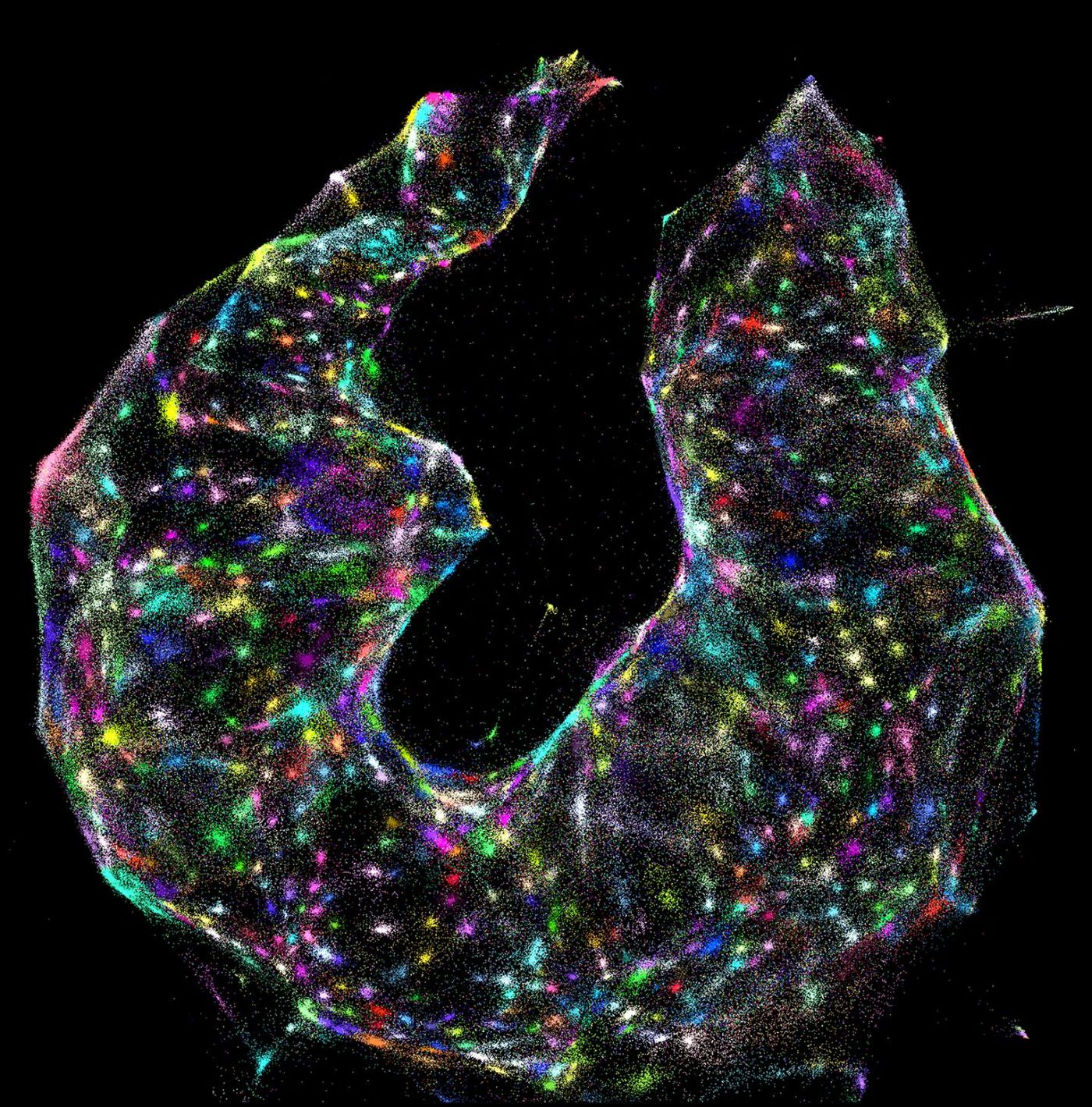
Ultimately, a two- or three-dimensional image is made that illustrates the position of molecules in space. A drawback is the difficulty in dealing with empty spaces, which the researchers are working to improve.
"We believe that the most exciting applications of this technology are in areas of biology in which mutations, RNA editing, and other forms of nucleotide-level variation work hand in hand within the organism to either produce physiological outcomes or cause disease," said biophysicist Joshua Weinstein.
In biological research, it’s critical to understand what happens on a cellular level. There are a variety of techniques for doing that, but many involve intense preparations and expensive equipment. It can also be difficult to see what many different molecules are doing at once. Now scientists can get a look at what happens in the cell on a genetic level.
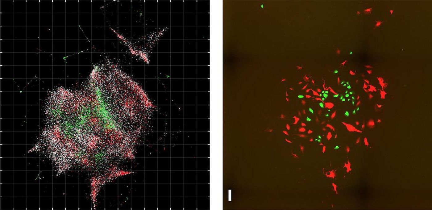
"You're basically able to reconstruct exactly what you see under a light microscope," Weinstein noted. He added that the techniques compliment each other; sparse molecules can be seen with light microscopy and dense molecules, even in clumps, can be resolved with DNA microscopy.
"By capturing information directly from the molecules being studied, DNA microscopy opens up a new way of connecting genotype to phenotype,” said HHMI investigator and molecular biologist Feng Zhang.
Weinstein discusses the technqiue in the video.
Sources: AAAS/Eurekalert! via HHMI, Cell Press, Cell