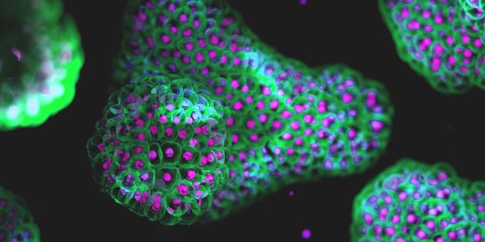
As cells grow into tissues in a developing embryo, they form sheets that fold to create the proper structures. The process follows a pattern that repeats as a member of a species grows, creating generally uniform results. Now, researchers at the Massachusetts Institute of Technology (MIT) studying the process in fruit flies have found that a network of proteins helps orchestrate the action, and builds redundancies into the system to create many alternative pathways that make the same thing. Their work has been reported in Developmental Cell.
In biology, morphogenesis is the process that gives an organism its shape. Embryonic cells join together into flat sheets of tissue, which fold into three-dimensional structures to create tubes, organs and many other parts of the body. If there is damage in some of those embryonic cells, an organism will still develop the proper shape.
“What we found is that there’s a lot of redundancy in the network. The cells are interacting and connecting with each other mechanically, but you don’t see individual cells taking on an all-important role. This means that if one cell gets damaged, other cells can still connect to disparate parts of the tissue,” explained the senior author of the study Adam Martin, an MIT associate professor of biology.
In this work, the scientists focused on gastrulation, a very early part of embryonic development that is similar in many organisms, including fruit flies and humans. Proteins called myosin and actin help form connections between cells, creating a tissue network. The researchers hypothesized that this cellular network influences the tissue folds.
“This is a process that’s fairly reproducible, and so we wanted to know what makes it so robust,” said study author and graduate student Pearson Miller.
The Martin lab teamed up with Jörn Dunkel, an MIT associate professor of physical applied mathematics to use an algorithm that can also assess the structure of galaxies. The computational tool identifies features of a three-dimensional structure; the team could trace networks of actomyosin through cells in tissue.
“Once you have the network, you can apply standard methods from network analysis — the same kind of analysis that you would apply to streets or other transport networks, or the blood circulation network, or any other form of network,” Dunkel said.
The team found many backups in the network, redundancies that protect the outcome if there is a problem in the process. Cells can find a way to fold correctly even if there is dysfunction in some areas. Their connections tend to line up with a furrow that is created in the early stages.
“We think this is setting up a frame around which the tissue will adopt its shape,” noted Martin. “If you prevent the directionality of the connections, then what happens is you can still get folding but it will fold along the wrong axis.”
The researchers want to follow up on this work in mice next. In the early parts of vertebrate development, folding occurs in the neural tube, which gives rise to the brain and spinal cord. When the tube fails to properly close it can lead to birth defects.
“We would like to understand how it goes wrong,” said Martin. “It’s still not clear whether it’s the sealing up of the tube that’s problematic or whether there are defects in the folding process.”
Sources: MIT, Developmental Cell