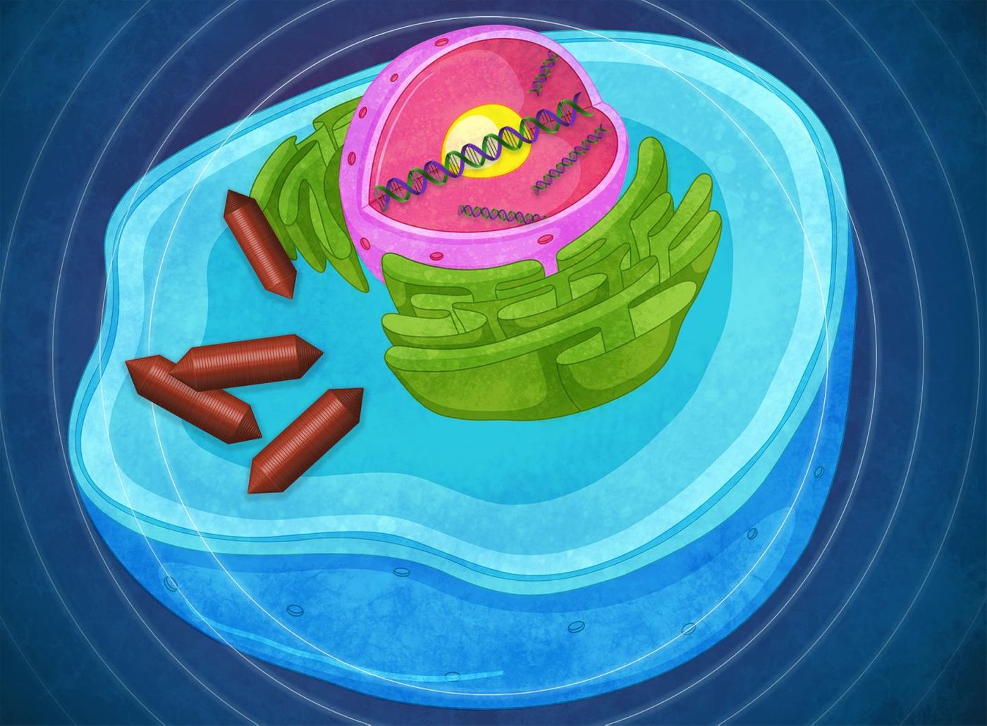
Understanding how and when and why certain genes are or are not active is a critical aspect of biomedical research. To learn more about gene expression, researchers often employ a fluorescent tag that can be added to the end of a gene sequence to create a type of reporter or marker gene. When a marker gene is expressed, the protein that is generated from that gene sequence glows, allowing researchers to see the expression in action. A very common fluorescent tag that is used to mark active genes is green fluorescent protein (GFP).
There are obvious limitations to this tool. Most organisms are not transparent enough to see the inside of, so if these fluorescent proteins are expressed deep within tissues, they can’t be seen until that tissue is processed or cut up. Now researchers at Caltech led by Mikhail Shapiro have engineered a type of gene reporter that can be seen with an ultrasound, which can penetrate tissue and reveal gene expression; they call it an acoustic reporter gene. The work has been reported in Science.
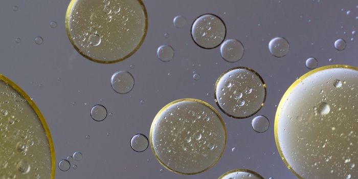
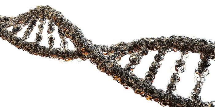
There is a species of bacteria that create tiny compartments filled with air and protein, called gas vesicles that can be seen with an ultrasound. Shapiro, graduate candidate Arash Farhadi and colleagues wanted to induce mammalian cells to create these bacterial vesicles, and to do so, they would have to transfer nine bacterial genes into a human cell line. However, the cellular machinery of bacterial and mammalian cells translates DNA in different ways.
"The translation machinery is very different in the two kinds of cells," Farhadi explained. "One of the biggest differences is that in bacteria it is common to have multiple genes arranged in the DNA such that they are transcribed into one shared piece of RNA, which is then translated into all the corresponding proteins, whereas in eukaryotes, every gene is usually on its own."
The researchers turned to viruses for a solution. "Viruses also need to trick mammalian cells into expressing a bunch of proteins," Shapiro said. "So we used viral elements to trick the cell into producing multiple genes from a shared piece of RNA." In the end, eight genes were included on one piece of RNA.
When the bacterial DNA was added to mammalian cells, the proteins found in gas vesicles were being generated, but not the vesicles themselves. The researchers found that the proteins would first have to be made in the right ratios.
"The correct ratios of proteins are programmed into the bacterial gene clusters, but when we put them into the mammalian cells, we have to figure out what those ratios need to be and how to get the mammalian cells to make those correctly," Farhadi said. It took years of work to find out what those ratios were, and now the vesicles are finally also being produced.
Shapiro and Farhadi will now be able to assess gene expression in a variety of structures, including tumors, neurons, immune cells, and many other kinds of cells. They are hopeful that other researchers will employ this technique on both research animals and maybe eventually humans in the clinic.
"There has been more than 20 years of work improving fluorescent proteins, and we probably have 20 years of work to improve what we've developed, but this is a key proof of concept," Shapiro added.
Experienced research scientist and technical expert with authorships on over 30 peer-reviewed publications, traveler to over 70 countries, published photographer and internationally-exhibited painter, volunteer trained in disaster-response, CPR and DV counseling.