Our cells contain a network of filaments called microtubules. Motor proteins, including one group called kinesins, move along them, ferrying cargo to different parts of the cell. Now researchers have used a cutting edge imaging tool to learn more about what different types of motor proteins carry. The findings are changing what we know about trafficking in cells. The work, reported in the journal Traffic, has provided new insights into the control of this movement in neurons, and shed new light on neurodegenerative disease.
"What we see is that there must be some other regulation beyond what is known that is directing kinesin motor proteins within a cell," said Marvin Bentley, assistant professor of biological sciences and member of the Center for Biotechnology and Interdisciplinary Studies at Rensselaer Polytechnic Institute. "There's a mysterious interaction, and we don't know what it is."
Human cells express 45 types of kinesin motor proteins. These molecules are structured like a pair of legs with a tail and carry many critical types of cellular molecules. Between fifteen and twenty kinesins use compartments called vesicles to move these materials. Researchers wanted to know more about how the kinesins are directed to carry the right stuff to the proper location.
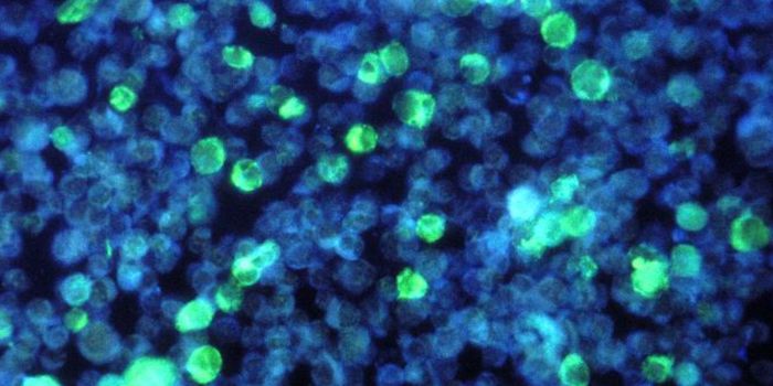
Fluorescent tags can be added to molecules in cells using transgenes or antibodies so their movement can be observed. A widely used tag is green fluorescent protein (GFP). However, tagging motor proteins this way often only generates a massive jumble of green; there are so many motor proteins, making it challenging to see the movement of individual molecules.
Previous work has tackled this problem by only tagging the legs, or motor domain, to see where they went. This research resulted in the “smart motor” theory, which suggests that the motor domain only travels on certain microtubules, and ferries the cargo that sits on the ends of those microtubules. Work by Bentley’s team has suggested that other influences are at work, however.
His team created a way to tag only the tail end of a kinesin. These tails attach to specific vesicles, and their tool illustrates that kinesins carry certain cargo. A few hours after a kinesin is expressed and before too many green tails are present, the researchers could identify the vesicles that were attaching to specific kinesins. The “smart motor” theory isn’t doesn't always explain this movement. Some kinesins aren’t found in the location indicated by the theory, and others were found in places they wouldn’t theoretically be able to get to. Vesicles that were attached to kinesins that moved to portions of neurons called axons or dendrites did not line up with what was preferred by their motor domain.
"It's clear that it's not solely the preference of the motor domain that determines where these things go, there's something else that's regulating them," noted Bentley. He suggested that adapter proteins play a role too, and is hopeful that additional research will elucidate the mechanism further.
"The movement of essential neurotransmitters and metabolites within a cell is amazingly complex, but we must understand these fundamental processes before we can control diseases where this cellular traffic has gone awry, particularly in neurons," said Curt Breneman, dean of the School of Science. "Marvin's inventive approach to this problem has yielded a fascinating insight and a new direction for further research."
Experienced research scientist and technical expert with authorships on over 30 peer-reviewed publications, traveler to over 70 countries, published photographer and internationally-exhibited painter, volunteer trained in disaster-response, CPR and DV counseling.