There are three types of muscle tissue in the human body including cardiac muscle, smooth muscle, and skeletal muscle. Skeletal muscle fibers are those which are attached to the skeleton. They are characterized as having striations (stripes) and are under voluntary control. For example, our brain tells us when to move our arm and contract the muscle – this is voluntary. Smooth muscle fibers are non-striated and makeup the walls of out organs, excluding the heart. Smooth muscles are under involuntary control. Cardiac muscle can be found only in the heart, is striated and is also under involuntary control.
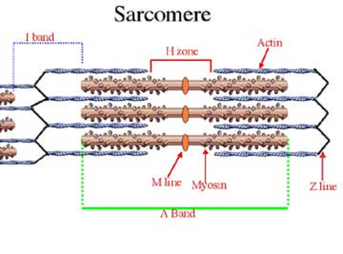
Muscles are made up of bundles of muscle fibers known as myofibrils. Myofibrils are made up of two different types of filaments known as actin and myosin. The myofibrils are organized into units called sarcomeres which give them the appearance of striations. The sarcomeres, which are the contractile units of the muscle, are made up of several distinct regions which are discernable by electron microscopy. The ends of the sarcomere are defined as the Z-disc (also known as the Z-line). Within the sarcomere there are light bands that alternate with dark bands. The dark bands are made of myosin filaments that are known as A bands. The light bands are made of actin filaments and are known as I bands. The myosin and actin filaments overlap in the peripheral regions of the A band where the middle region contains only myosin. This middle region is known as the H zone. The actin filaments are attached at their ends to the Z disk where crosslinking of protein α-actinin occurs.
Researchers from London used
electron tomography and
subtomogram averaging to visualize the lattice-like three-dimensional structure of the Z-band in cardiac muscle in mice. The muscle Z-band connects antiparallel actin filaments from two adjacent sarcomeres. Tension builds up via transmission though α-actinin linking of the antiparallel filaments. Through their research, scientists discovered that the Z-band is made up of overlapping ends of actin filaments crosslinked by 4-6 layers of α-actinin. The Z-band also has a terminal actin filament of 5-7 nm that is devoid of links and is possibly capped by a protein known as CapZ.
The identification of α-actinin and the CapZ terminal protein of the Z-band is significant considering that α-actinin mutations have been linked to cardiac disease including hypertrophic cardiomyopathy and arrhythmias. In addition, previous research has shown that CapZ helps protect against damage caused by ischemia (lack of oxygen). Further study of the Z-band topography may help determine how to prevent mutations in α-actinin and CapZ to prevent or treat cardiac diseases such as myopathies.
Sources:
Journal of Molecular Biology;
US National Library of Medicine;
The Cell: A Molecular Approach;
Fernando Amat Bioimaging Research;
American Society for Microbiology