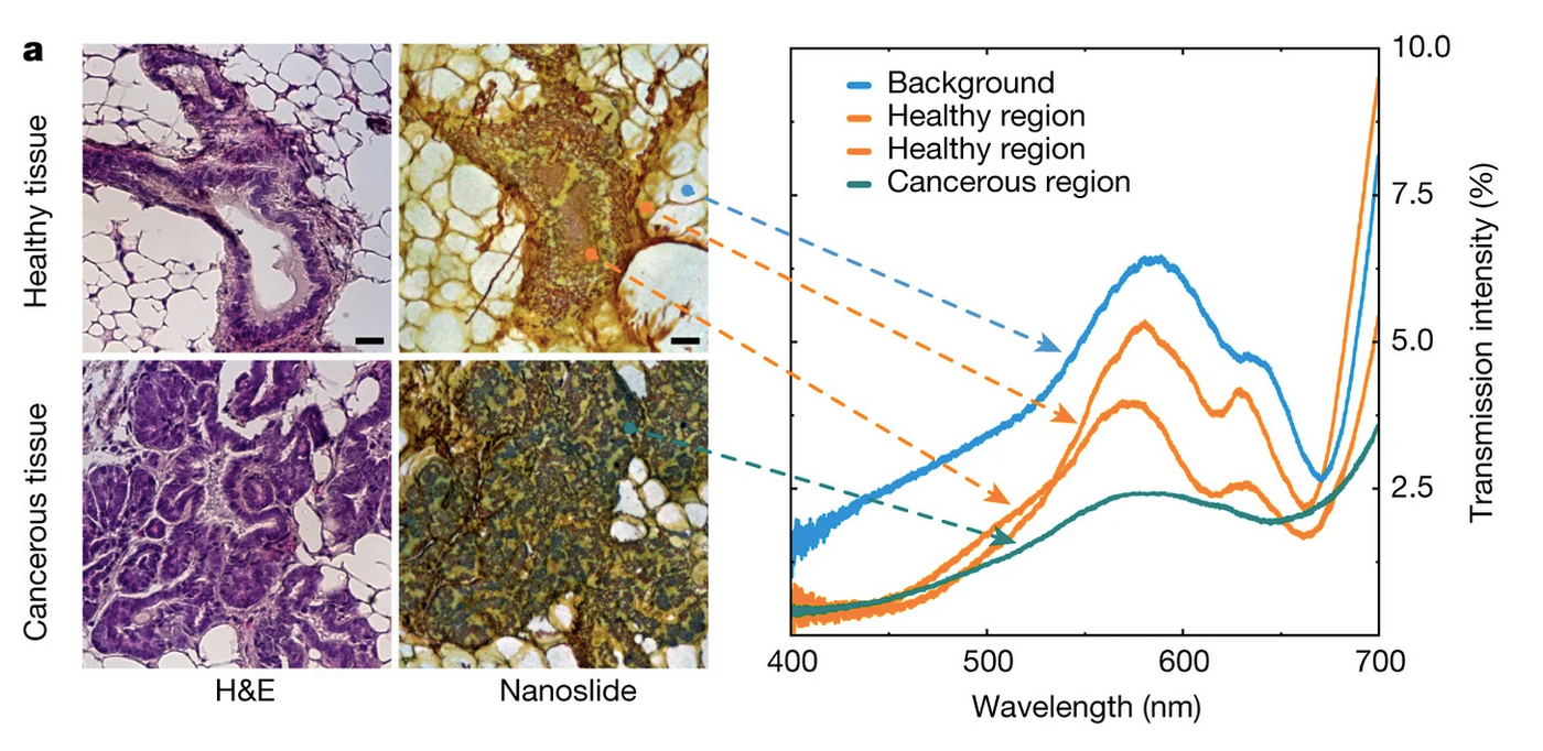
When cancer is diagnosed, a sample has to be taken from a patient, treated, and analyzed. Researchers have now modified this method slightly to improve detection. Biopsies are assessed on glass slides, and a new type of glass slide has been created called NanoMslide, which can cause a color change to indicate when cancer cells are present. The work has been reported in Nature.
The standard approaches in tissue analysis typically involve applying a stain or immunological label to a sample. But the results aren't always easy to interpret, and there is a "risk that some samples are misdiagnosed, particularly during the very early stages of disease," said one of the project leaders, Professor Brian Abbey.
"Recent breakthroughs in nanotechnology have allowed us to manipulate the interaction of light with biological tissue so that abnormal cells appear to have a different color to healthy ones. Comparing images from our slides to conventional staining is like watching color television when all you've seen before is black and white."
These slides have a nanofabricated coating, which turns a glass slide into a sensor. It only takes light to activate that sensor, and when something like tissue touches the sensor, an array of colors are produced; tissues can be see in a spectrum of color when thin slices are placed on this type of slide. When the researchers tested various types of tissues on the NanoMslide, they found that certain colors would routinely indicate when cancer cells were present.
With current tools, it can be hard to tell the difference between benign lesions and the early stages of breast cancer, noted another study leader, Associate Professor Belinda Parker. If there are only a small number of abnormal cells, it can be especially challenging. The researchers suggested that NanoMslides will make these diagnoses easier.
"When I first looked at a tissue under the microscope on the NanoMslide, I was incredibly excited," said Parker. "For the first time I saw cancer cells just popping up at me. They were a different color from the surrounding tissue, and it was very easy to distinguish them from surrounding cells."
Parker suggested that NanoMslides could be used with current staining protocols to make more accurate diagnoses. "Based on our preliminary findings with the NanoMslide, we think this platform could be really useful in early breast cancer diagnosis, but also in other cancers where we're really just trying to pick up a few cancer cells in a complex tissue or a blood sample."
Experienced research scientist and technical expert with authorships on over 30 peer-reviewed publications, traveler to over 70 countries, published photographer and internationally-exhibited painter, volunteer trained in disaster-response, CPR and DV counseling.