 3D cell cultures are no longer a futuristic idea. They’re already reshaping how we study diseases like cancer, offering more realistic models of how cells behave in the body.
But despite their advantages, 3D workflows have a reputation for being complex, labor-intensive, and hard to scale.
That’s starting to change. With better workflow design and integrated tools, building and analyzing 3D cancer models is becoming almost as straightforward as working with 2D cultures.
Let’s look at how automation and live-cell analysis are helping make 3D workflows more accessible for cancer research.
Why Simplifying 3D Workflows Matters
For all their advantages, implementing 3D workflows in routine drug discovery comes with several persistent challenges:
- Manual Matrix Handling
- Preparing and dispensing hydrogels by hand is slow and inconsistent. Even small variations in gel composition or volume can impact how cells grow, and how reliable your results are.
- Reproducibility Gaps
- Especially with biologically derived materials, batch-to-batch differences happen, making it hard to compare results across experiments or labs.
- Imaging Limitations
- Traditional plate readers or endpoint assays often miss the full picture. Many rely on invasive, destructive labeling methods. And when it comes to opaque or curved 3D structures, standard imaging systems can struggle.
- Throughput Bottlenecks
- 3D workflows tend to need more hands-on time, specialized materials, and extra steps, things that clash with the fast-paced, high-volume demands of drug discovery.
It’s no wonder some research teams hesitate to make the switch, especially in time-sensitive fields like oncology.
An Integrated Approach to 3D Culture and Analysis
Fortunately, recent advances are making it much easier to overcome these hurdles. Take, for example, the combination of the RASTRUM™ 3D cell culture platform and the Incucyte® live-cell system — together, they offer a streamlined, scalable workflow designed with scientists in mind.
1. Reproducible 3D Model Generation
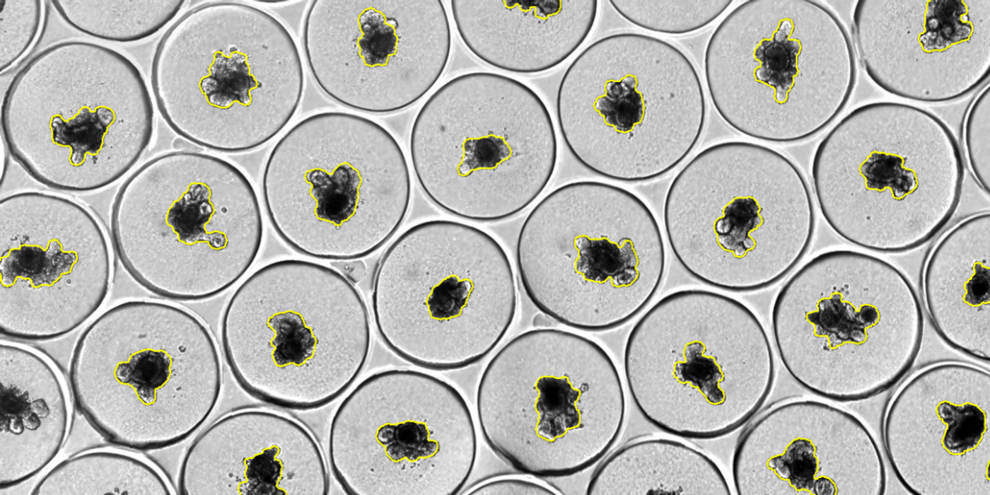
In this workflow, 3D cultures are produced by bioprinting cell suspensions into synthetic, tissue-like matrices within standard 96-well plates. This approach with the RASTRUM™ 3D platform reduces the need for manual gel preparation and helps ensure greater consistency across experiments.
The stiffness of these matrices can be tuned, and they are functionalized with extracellular matrix peptides, enabling researchers to better replicate aspects of the tumor microenvironment and study how it influences cell behavior.
2. Live-Cell Imaging Without the Guesswork
Once the 3D cultures are established, they can be monitored continuously using brightfield imaging on the Incucyte® system. This allows for real-time observation of spheroid growth, cell viability, and treatment response, without requiring disruptive labels or endpoint staining.
3. Integrated Data Analysis Made Simple
The same workflow includes tools for automated image analysis, generating growth curves, morphological data, and dose-response information with minimal manual processing. This streamlines the path from image acquisition to biological insight, helping researchers interpret their data quickly and consistently.
Case Study: Breast Cancer Cells in Tunable 3D Matrices
A recent collaboration between Sartorius and Inventia Life Science, a biotech company specializing in 3D cell culture, highlights the power of these tools in action.
In the study, researchers explored how matrix stiffness influences cancer cell behavior by culturing MCF-7 breast cancer cells in RASTRUM™ matrices at three different stiffness levels: 0.7, 1.1, and 3.0 kPa.
Over 10 days, they monitored the cultures with live-cell imaging to track growth and treatment response in real time.
Key Findings:
- Softer matrices (0.7 kPa) supported steady, linear growth
- Stiffer matrices (1.1 and 3.0 kPa) showed growth plateauing after day 8
- The differences were statistically significant (p < 0.05)
This revealed a clear window of active growth, crucial information for optimizing when to introduce treatments.
When the same cultures were exposed to chemotherapeutic agents like camptothecin, 5-FU, and staurosporine, researchers could see time-dependent cytotoxic effects as early as 1.75 days after treatment. By day 10, the dose-response trends were clear and robust.
Explore the full case study here.
Why It Matters
This integrated, automated workflow tackles some of the biggest barriers in 3D assay development:
- Better Reproducibility — Automated dispensing minimizes variability
- Higher Throughput — Standard formats and bioprinting support scalable screening
- Real-Time Insight — Non-invasive imaging provides kinetic data without disturbing the cultures
“What makes this workflow stand out is how reproducible and scalable it is. You can run dozens of plates with confidence that the data will be consistent across every well.” — Dr. Sean Porazinski, Application Science Lead, Inventia Life Science
Making Better Biology More Accessible
3D models are quickly becoming the gold standard for preclinical cancer research. But for widespread adoption, they have to be practical, not just scientifically powerful.
Streamlined solutions like RASTRUM™ and Incucyte® are helping research teams move from culture setup to actionable insight faster, without compromising data quality.
If your lab has been hesitant about adopting 3D workflows, now might be the perfect time to take another look. With the right tools in place, modeling complex biology doesn’t have to mean complex processes.
“We’re seeing researchers use this workflow to uncover not only how cells grow in different mechanical environments, but also how they respond dynamically to compounds. It’s a step forward in making complex models more accessible and usable for everyday drug screening.” — Dr. Sean Porazinski, Application Science Lead, Inventia Life Science
Read the detailed case study here.