There is a huge amount of DNA in most human cells, and that DNA has to be carefully compacted and organized so that it will fit into a cell’s nucleus, while the crucial parts of it remain accessible so that genes can be expressed. DNA is carefully arranged and wrapped around proteins, forming structures known as nucleosomes, which look a bit like beads on a string. These beaded strings are then compacted further to make chromatin fibers, which fit into the nucleus. Now scientists have learned more about this process, and have used powerful tools to visualize it. The work has been reported in Science.
In cells, droplets known as condensates can form. Like oil droplets in water, these condensates can bring molecules together and carve out a space that is a bit separated from the rest of the stuff in the cell. Previous work has shown that nucleosomes made in the lab will migrate into condensates.
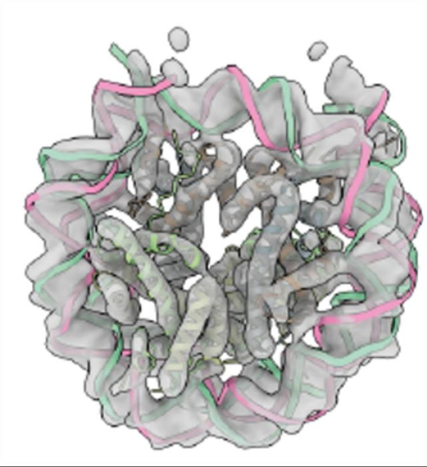
With imaging tools, researchers were able to capture images of synthetic chromatin condensates. This work showed how chromatin fibers are packaged in these condensates. The investigators also used computational tools and additional microscopy work to determine how molecules were interacting inside of the condensates.
They found that the length of DNA molecules that linked the nucleosomes together also affected their three-dimensional arrangement, how chromatin interacts, and the condensate structure.
These findings can explain why different types of chromatin can create different material characteristics, and why some chromatin is better at phase separation than others.
“The work has allowed us to tie the structures of individual molecules to macroscopic properties of their condensates, really for the first time,” said senior study author and Howard Hughes Medical Institute (HHMI) scientist Michael K. Rosen of The University of Texas Southwestern Medical Center. “I’m certain that we’re only at the tip of the iceberg–that we and others will come up with even better ways of developing those structure-function relationships at the meso scale.”
This study can also help scientists use these methods to understand other condensates, structures that we still have a lot to learn about. Since condensate formation goes awry in some diseases, this work can also advance the study of disease.
“By doing this research, we will better understand how abnormal condensation could lead to different diseases and, potentially, that could help us develop a new generation of therapeutics,” explained first study author Huabin Zhou, a postdoctoral scientist in the Rosen Lab.
Experienced research scientist and technical expert with authorships on over 30 peer-reviewed publications, traveler to over 70 countries, published photographer and internationally-exhibited painter, volunteer trained in disaster-response, CPR and DV counseling.