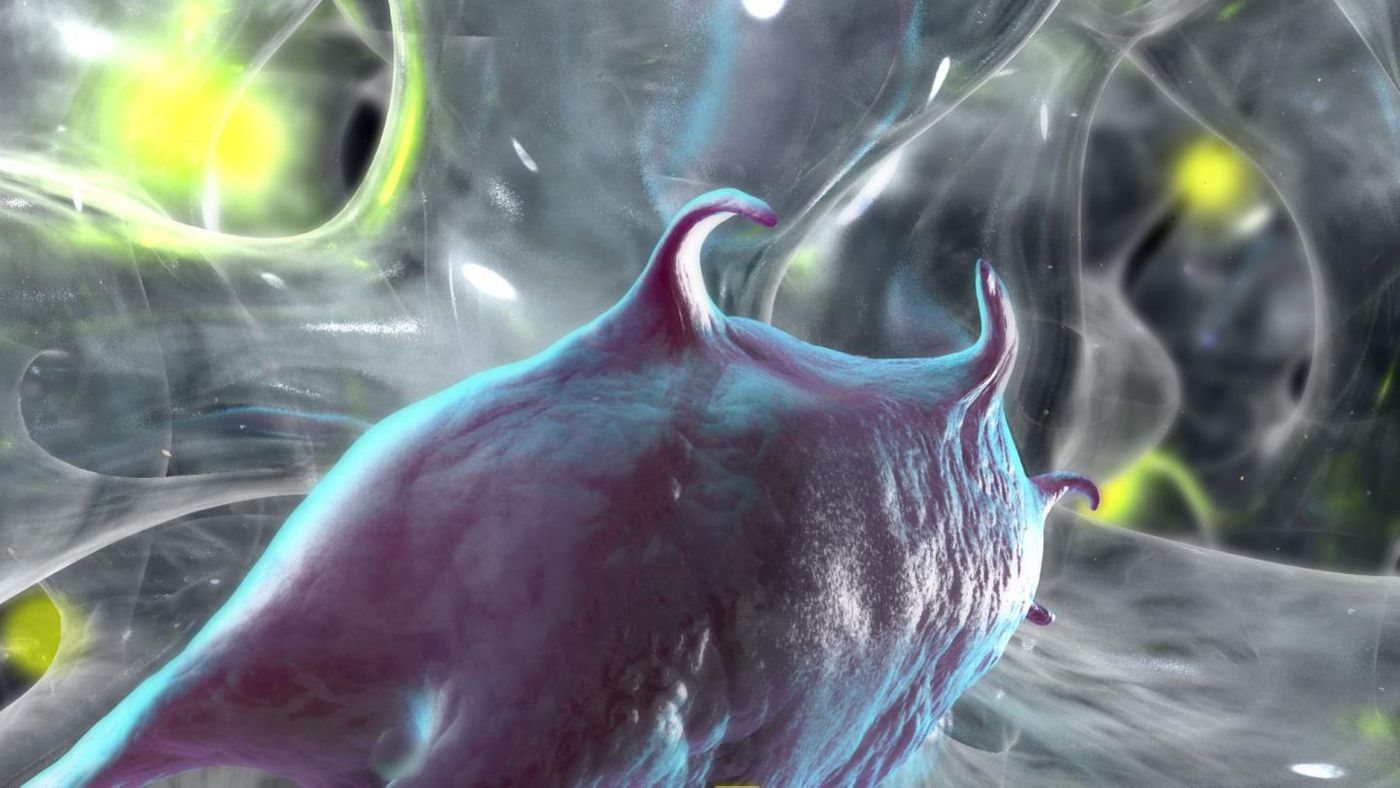
If we are ever to use stem cells in therapeutics and regenerative medicine, there must be a complete understanding of their processes and properties. One such process is how stem cells interact with their environment.
"Cells do not simply reside within a material, they actively reengineer it," explained Kelly Schultz, an Assistant Professor of Chemical and Biomolecular Engineering at Lehigh University's P.C. Rossin College of Engineering and Applied Science. "Characterizing how cells behave in 3-Dimensional (3D) synthetic material is critical to advancing biomaterial design used in wound healing, tissue engineering and stem cell expansion."
Schultz has shown in
work published in the Proceedings of the National Academy of Sciences that cells degrade stuff in the area around it during attachment, spreading and movement, in a way that was completely different from what had been thought. Scientists had thought that as cells move through a material, they degrade it at the same time.
The investigators upended this theory using a technique called microrheology. Stem cells were encapsulated in a hydrogel, and then the researchers measured the changing interactions between cells as they moved through the gel.
Surprisingly, the scientists observed that the cells paused before moving. At a so-called Point A, a cell secretes an enzyme. The scientists suggest that the enzyme is then stuck to an inhibitor that halts any degrading actions of the enzyme as the cell moves to a Point B. At that Point B, the enzyme or enzymes then digest the material surrounding the cell. The cell then moves on to the next location. The previously mentioned inhibitor also functions to prevent enzymatic degradation of material as the enzyme is being secreted.
In research the team plans to carry out, the inhibitor, or combination of molecules that creates the inhibition, will be identified and characterized such that it can be controlled. Cells release four kinds of enzyme inhibitors; they are known as tissue inhibitor of metalloproteinases. The investigators hope that identifying how they shut off the enzymatic degradation will allow them to shut off the inhibitor during secretion, speeding up cell movement by consequence.
Schultz explains that quicker cell motility has an impact on wound healing - the faster cells arrive at the site of injury, the sooner the repair work can start. "Our goal is to prevent the cell from stalling, encouraging it to become active right away and arrive at the wound site at twice the speed," she said. Check out the video below for animation of their work.
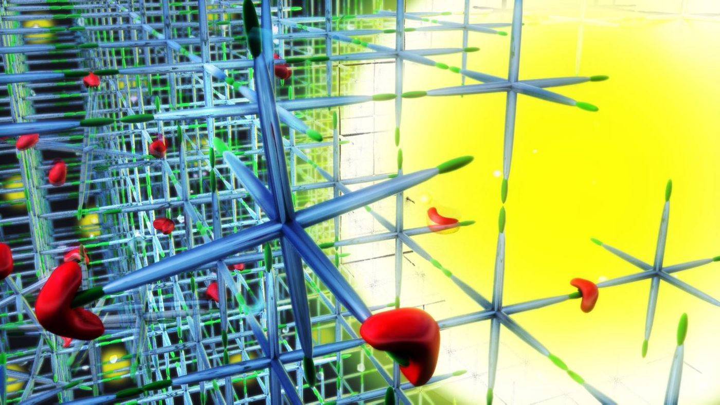
In their work, the researchers utilize hydrogel for a couple of reasons. It is a good replica of tissue, providing an excellent physical matrix by which 3D cell movement can be studied. Hydrogels can mimic a variety of tissues and can be specialized to replicate various properties of different cells. They can also be implanted into people and used as a scaffold; especially on large wounds, that structure can be critical to successful healing.
"For example, if a patient is missing a piece of bone that is too large to heal - or regenerate - on its own, a hydrogel structure can be implanted. Inside the structure are stem cells that have been given an environment that would push them down a lineage into bone cells," Schultz explained. "Once implanted the stem cells reproduce themselves, 'filling in' the part of the bone that is missing. While the bone is growing outside the implant, new tissue is growing on the inside--speeding healing. As the bone regenerates, the implant disintegrates."
Schultz mixes a variety of ingredients from polymers to stem cells and microparticles to create the hydrogel structures. During the enzymatic degradation, microparticles allow the hydrogel’s physical properties to be measured.
"We take videos of the microparticles and observe their movement," explained Schultz. "The amount they wiggle reveals their properties and enables us to see spatially what's occurring. That is how we characterized the unexpected degradation profile."
While this work focuses on the remodeling of synthetic scaffolds by human mesenchymal stem cells, the methods under development are expected to broaden our understanding of the interactions of cells and materials as well as testing how best to engineer cellular behavior for tissue regeneration applications and 3D cell culture protocols.
Sources:
AAAS/Eurekalert! via
Lehigh University,
Proceedings of the National Academy of Sciences