The production of two cells from one, cell division, is one of the core functions in life. Cells divide many times during development and as the cell number increases, the cell types become more specialized and organized. The number of times a fertilized egg divides and the position of the daughter cells can be a very important factor in the ultimate form of the organism.
While much is known about this process, one that is highly conserved through evolution, how the orientation of cell division affects the body axis of an animal is not known.
New work published in the journal eLife shows that in an organism called a sea squirt, seen in the video above, a special structure in the cell membrane orchestrates the orientation of cell division machinery in epithelial cells of the embryo. Sea squirts were selected for this study because with only a small number of cells, they present a simple model for the study of cell division.
The first author of the study, Dr. Takefumi Negishi, commented "When I observed this structure dynamically moving and extending within the cell, I immediately thought this might be playing an essential role for cellular function." The structure was called an invagination.
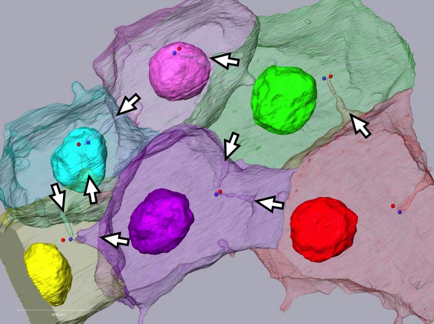
Using fluorescence microscopy, live embryos were imaged undergoing their final cell division, in which most of the cells are dividing in the same direction - shown in the video (in which ten minutes have been compressed to one second). Results showed that the posterior or rear end of each cell gets pulled into the cell, forming those invaginations. The invaginations then elongate intracellularly in the direction of the centrosome, which is a critical part of the cell division machinery. The centrosome gets pulled toward the posterior end of the cell, where it then duplicates. The daughter centrosomes subsequently line up with the invagination, and a spindle then forms.
The investigators used an advanced type of electron microscopy called Serial block-face (SBF) scanning electron microscopy (SEM) – SBF SEM. For a demonstration of how this technique works, check out the video below. An ultramicrotome slices thin sections off of a block of sample, then the freshly revelaed face is imaged. This is repeated many times to generate a 3D reconstruction of the sample.
The authors thus suggest a completely new model for controlling cell division orientation, one in which the polarized membrane structure repositions the cell division machinery at a specific end of the cell, impacting the orientation of subsequent cell division.
One supervisor of the study, Professor Naoto Ueno of the National Institutes of Natural Sciences in Okazaki, Japan, said, "We hope our findings facilitates studies on similar membrane structures in other animals that might have diverse biological functions."
Sources:
AAAS/Eurekalert! via
National Institute for Basic Biology,
eLife