When the molecular components of a cell are under study, subcellular nanomachines are often analyzed in isolation from the remainder of the cell because of the limitations of tools that are available for use. It is difficult to visualize structures smaller than a cell as they perform their functions, and it is very challenging to obtain the resolution to see structural changes in proteins are they move in live cells. Publishing in Cell, scientists have now managed to visualize the function of a cellular structure called the exocyst.
"The in vitro techniques available are excellent and allow us to make observations at the atomic level, but the information provided is limited. We will not know how an engine works if we dissemble it and only look at the individual parts. We need to see the engine assembled in the car and running. In biology, we still do not have the tools to observe the inner workings of a living cell, but the technique that we have developed is a step in the right direction and we can now see, in 3D, how the protein complexes carry out their functions," explained the leader of this work, Oriol Gallego of IRB Barcelona.
In order to get a look at cellular structures in live cells, the researchers took advantage of the power of Nobel-prize-winning super-resolution microscopy techniques, combining them with cell engineering and computation. These tools enabled the study of protein complexes at a resolution of 5 nm, which is "four times better than that offered by super-resolution and that allows us to perform cell biology studies that were previously unfeasible," explained Gallego.
Cells were engineered to create artificial supports that would anchor protein complexes. The support designed enabled the investigators to manipulate the angle from which the stationary nanomachinery is studied. To figure out the three-dimensional protein complex structure, the super-resolution microscopy was used to calculate the distances separating various complex components, finally combining all of the information.
One function of exocytosis, which expels material from the cell, is to send signals to the outside of the cell. Neurons for example, release neurotransmitters by exocytosis, and those neurotransmitters are picked up by neighboring neurons. In this work, the researchers have determined the complete structure of a mysterious nanomachine important to exocytosis. "We now know how this machinery, which is formed by eight proteins, works and what each protein is important for. This knowledge will help us to better understand the involvement of exocytosis in cancer and metastasis - processes in which this nanomachinery is altered," he explained.
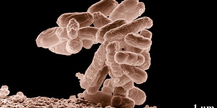
This method can be extrapolated to study dysfunction in cellular processes that leads to disease. Viruses and microbes can now be visualized commandeering cellular nanomachinery during infection. Knowing what happens at the molecular level during illness could lead to improved therapeutics.
Relatively large protein structures can be studied in this way. "Being able to see protein complexes measuring 5 nm is a great achievement, but there is still a long way to go to be able to observe the inside of the cell at the atomic scale that in vitro techniques would allow," said Gallego. "But, I think that the future lies in integrating various methods and combining the power of each one.”
This project took Oriol Gallego five years to develop at the Molecular Medicine Program at IRB Barcelona. Gallego will continue pursuing this work in Japan and Germany. "After, I would like to continue to do top-level research in Barcelona, and I hope that this study that has been published in Cell helps me to do so," he said, in an aim to "make the invisible visible.”
Sources: AAAS/Eurekalert! via IRB Barcelona, Cell