Looking at the dynamics of living cells is very challenging compared to looking at cells that have been fixed in stasis, even though live cells are far more relevant to scientific study. As such, researchers have been working to find ways to use live, instead of fixed cells in microscopy. A new method has been developed by researchers at the University of Bielefeld in Germany, which allows such observation of structures in living cells.
The work has been published in the open access Nature journal
Scientific Reports. In it they demonstrate that their method of microinjecting cells with molecules, after which nine of ten cells are still surviving. “The method works very well on fixed, that is non-living cells,” commented Professor Dr. Thomas Huser, head of the Biomolecular Photonics research group. “However, the problem is that much of what we want to know can be gained only from living cells.”
Scientists have been working to overcome the hurdles of visualizing molecules and structures inside of living cells. Usually when a researcher wants to get a look at cellular materials, the cell has to be fixed so it can then be chemically manipulated with antibodies that bind to specific proteins or structures of interest, and then those antibodies are illuminated with fluorescent tags. With typical microscopy techniques, scientists are able to visualize what happens inside of cells only under specific conditions. “Living cells impede the intrusion of most fluorescent probes,” explained Dr. Simon Hennig, a physicist who is working in Huser's research group.
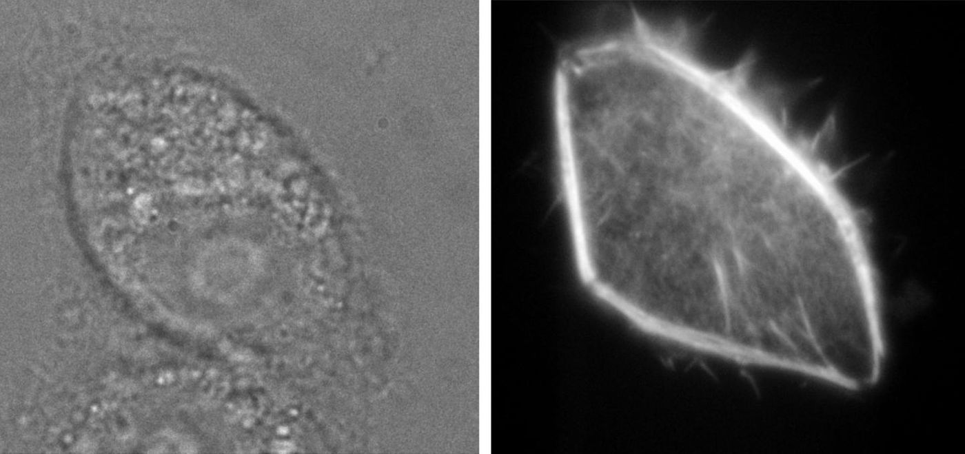
Their nanoinjection process involves using special, miniscule glass pipettes that send molecules right into live cells. A computer is used for this fine work, along with specialized instrumentation for insertion of the glass pipette into cells. Some people might be familiar with microinjection; this pipette is extremely small compared to the usual microinjection pipette sizes. This pipette is able to deliver only the molecule into the cell without any extra liquid, keeping the size of the cell constant.
“The method is so precise that we can even deliver the molecules to the nucleus of a cell,” said Hennig.
The researchers have confirmed that their protocol is useful and well tolerated across many different types of cells with myriad molecules. “This proof was necessary, because previous techniques such as microinjection harm the cells so much that most do not survive the treatment,” explained Hennig. Matthias Simonis, another study author, used their nanoinjection process on over 300 cells, and then compared what they saw to the products of a microinjection protocol. They saw that 92 percent of the cells had lived through the nanoinjection, while only 40 percent had made it through the microinjection process.
“The analyses also confirmed that these treated cells proliferated normally,” said Hennig. He suggested that proliferation doesn’t only indicate a healthy cell; it also opens the door for more investigation. Because negative impacts from the injection can be ruled out, researchers are then able to analyze the injected cells while eliminating artifacts or nonspecific effects that result only from the injection. Nanoinjection, Hennig said, could help scientists understand how cells interact with and react to one another.
If you are interested in learning more about standard fluorescence miscroscopy and how it is used in biomedical research, check out the video above featuring Dr. Nico Stuurman of UCSF.