A new technology developed by University of Central Florida’s Aristide Dogariu is an optical fiber light beam, used to monitor a patient’s blood in real-time during surgery. With constant, updated information on a patient’s blood status, surgeons can better prevent life-threatening blood clots during surgery.

"It provides continuous feedback for the surgeon to make a decision on medication," Dogariu explained. "That is what's new. Continuous, real-time monitoring is not available today. That is what our machine does, and in surgeries that can last for hours, this information can be critical.”
Why do doctors need to know the status of a surgical patient’s blood during a procedure? One of the main concerns during surgery of any kind is the chance of the patient’s blood clotting, or coagulating, too quickly, which can lead to a stroke or blood clot in the lungs.
It is particularly important to prevent blood clots during a procedure involving the heart, because clots can interfere with the heart-lung machine used to circulate blood in and out of the body. A surgical patient receives anticoagulation drugs before a procedure, but to make absolutely sure of the blood’s status, their blood is still tested every half an hour or so. Analysis takes about ten minutes, and given that some surgeries last more than four hours, there are several ten-minute periods where the status of the patient’s blood is unknown.
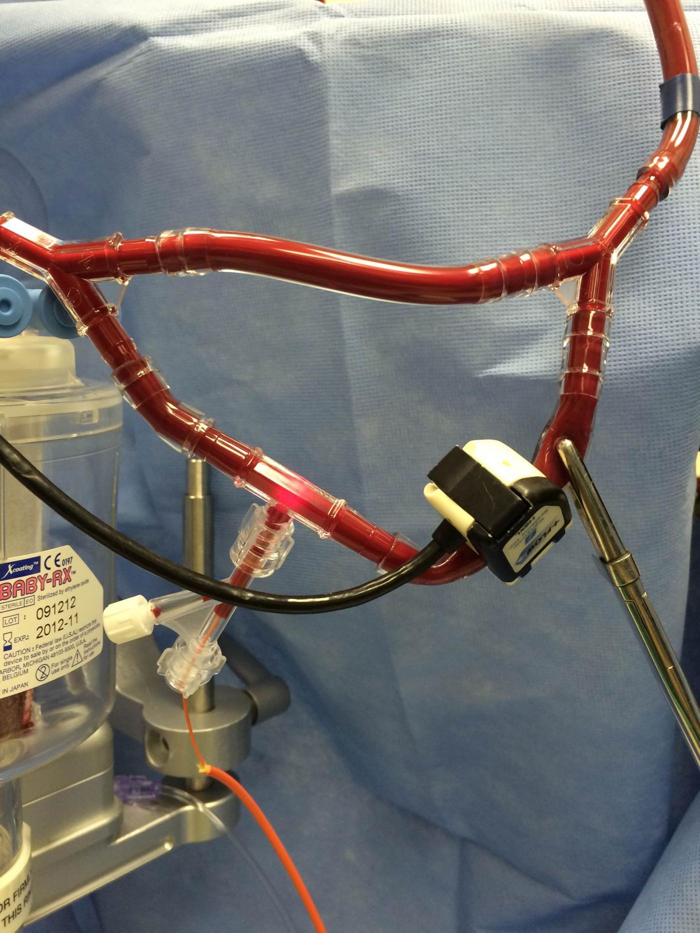
Dogariu’s new technology replaces the wait time for testing after a patient’s blood is drawn. The optical fiber light beam apparatus connects to the tubes of the heart-lung machine, providing constant status updates, so if the patient needs more anticoagulant drugs, the surgeon knows immediately.
The light beam technology has been tested during 10 cardiovascular procedures with infant patients over the past year, and each test was successful.
"I absolutely see the technique having potential in the intensive care setting, where it can be part of saving the lives of critically ill patients with all kinds of other disorders," said Dr. William DeCampli, chief of pediatric cardiac surgery at Arnold Palmer Hospital for Children, the leading center for pediatric cardiac surgery in Orlando and among the best nationwide.
The present study describing the new technology is published in the journal Nature Biomedical Engineering.
Source: University of Central Florida