To investigate the intricate processes underlying biology, scientists often have to take a close look at incredibly tiny structures, something that isn’t always very easy. There have been incredible advancements in microscopy since it was first developed several hundred years ago, and the technology continues to improve all the time. Researchers can now get an accurate look at stuff that exists on the nanoscale, at only a few billionths of a meter wide. When such small objects in biological tissue are under study, structures and proteins in the cell usually have to be labeled. That is often done with fluorescent tags or antibodies, that can be seen under specialized lighting in a microscope. There are many technical limitations to such work, however.
A new technique aims to address some of those hurdles. Cells in the brain are densely interconnected and closely related; to get a better look at those relationships, scientists have created SUSHI, or Super-resolution Shadow Imaging. It was developed by the team of Dr. Jan Tønnesen, a researcher in the Ramón y Cajal Programme at the UPV/EHU's Department of Neurosciences.
Reporting in Cell, the tool is specifically for imaging live brain tissue. Now a researcher does not have to label each cell of interest, but instead, fills the empty spaces in between in one fell swoop. The label also stays outside of the cells, acting as a negative contrast to the brain cells it surrounds. This process makes for a much easier and more straightforward way to see cells in living tissue.
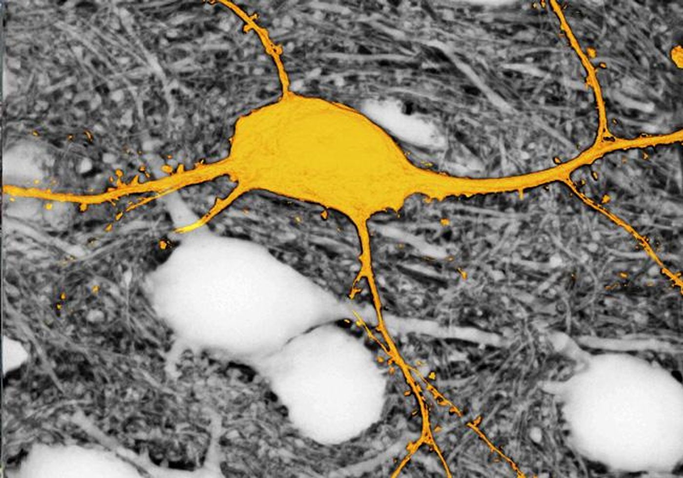
"The SUSHI technique is revolutionary because it allows us to simultaneously image all the brain cells in a specific region of living brain tissue,” noted Dr. Tønnesen. “In the past we used to come across blank spaces in the microscopy images, because we were unable to label all the cells at the same time. This fact was a big constraint for us. From now on, this technique will enable us to see all the cells in the area of study that we put under the microscope lens as well as all their interactions, and that will allow us to advance our knowledge of brain functions in a healthy organ and in a diseased one".
You can check out a video above of images of brain cells produced by the scientists. In the video below, you can hear more about super-resolution microscopy.
Sources: AAAS/Eurekalert! via University of the Basque Country, Cell