In the near future, the X-ray images from your doctor's office will no longer be just black and white, at least that's what MARS Bioimaging Ltd, an imaging company based in New Zealand, hopes. Why are they so confident? A CERN (yes the organization that built the Large Hadron Collider)-developed technology called Medipix3 that can capture a three-dimensional color x-ray picture of the human body.
Wait a minute, color is, in fact, a characteristic of light (or electromagnetic radiation to be scientifically accurate) as your common sense may alarm you. The color spectrum of visible light rays is way off from that of x-ray.
The visible light comes with the range of wavelengths humans can perceive approximately from 390 to 700 nm. Within it, red is somewhere between 700 and 635 nm, and blue at the other end between 490 and 450 nm. Yet, the spectrum of x-ray is nowhere close: from 0.01 to 10 nm.
So how can a "color" x-ray be possible? You can get a hint from an airport or security x-ray scanner. In those images, items of different densities are assigned with different colors by computing algorithms: blue and black suggest the material could be metal, hard plastics, or alloys; orange indicates organic or even biological materials like cotton fabrics, food, or explosives; and green is for thin plastics and rubber. In general, the denser the material, the more radiation gets absorbed and blocked.
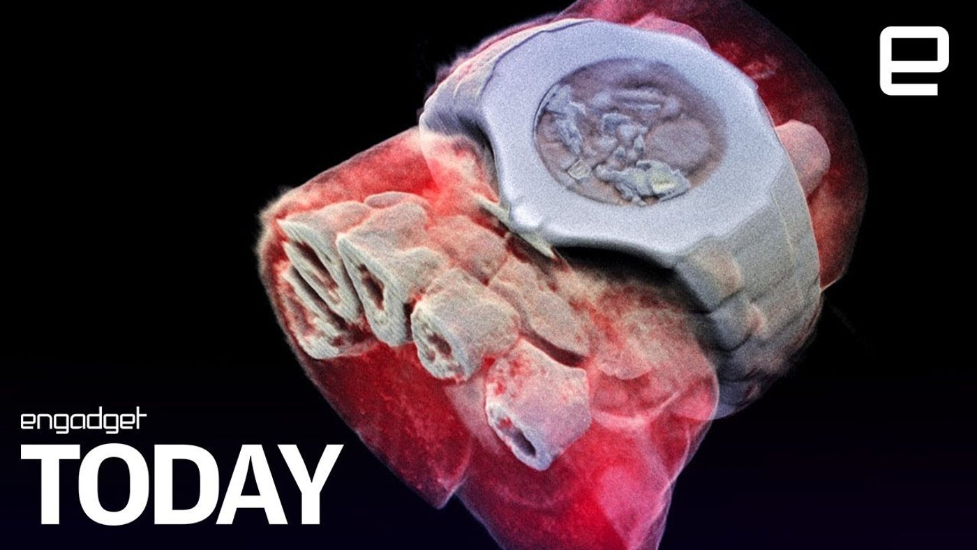
A similar principle applies in this color x-ray scanner. When x-rays of various spectra get beamed at a human subject, the Medipix chips which are "high-resolution, high-contrast" cameras capture individual photonic particle that goes through the body and hit their pixels. The electronic shutter allows the detector to identify photons of different energy level, whose energy signature enables the scanner to differentiate and assign colors to a variety of tissues such as muscle, fat, water, and bones. Measurements of the photons are then processed with computer algorithm for the construction of a 3-D color image.
Color is a powerful indicator. Compared to grayscale x-ray images, full-color images allow doctors to conduct more accurate diagnoses. When discussing the advantage of using Medipix3 chip in medical diagnostic imaging, Phil Butler a co-developer of the x-ray scanner and a scientist of MARS Bioimaging Ltd said: "Its small pixels and accurate energy resolution mean that this new imaging tool is able to get images that no other imaging tool can achieve".
The Medipix technology was initially designed for particle tracking purpose at the Large Hadron Collider. After two decades of work and improvement, the chips have demonstrated a great potential for applications outside of high-energy physics.
Preliminary studies have been conducted in oncology, bone and joint disorder, cardiovascular diseases using a smaller version of the new technology. Clinical trials of this revolutionary technology on rheumatology and orthopedic patients are expected to happen later this year.
Scientists develop 3D, full-color x-rays (Engadget)
Source: Engadget/CERN