During its annual member meeting in June the Society of Nuclear Medicine and Molecular Imaging, an international body that promotes the science, technology and excellence in practice of nuclear medicine and molecular imaging, awarded the 2017 image of the year to a team of German researchers. The work provided by the group is a pictorial summary of data and images that showcase the three-stage application of a dual-labeled imaging probe designed for prostate cancer diagnosis.
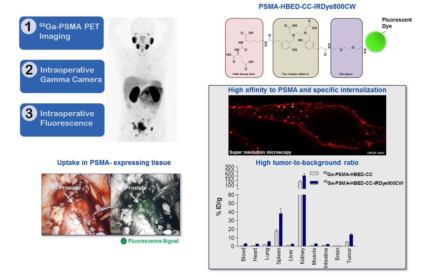
Dual-labeled PSMA-inhibitors for PET/CT imaging and fluorescence-guided intraoperative identification of prostate cancer and metastases. Credit: Society of Nuclear Medicine and Molecular Imaging
Prostate is a small walnut-shaped gland that produces the sperm-transporting seminal fluid. About one out of seven men will be diagnosed with prostate cancer during his lifetime. When detected early, 90% prostate cancer is curable. Because treatment is highly individualized, molecular imaging technologies are dramatically improving staging and treatment planning: it helps to pinpoint the location of a tumor, and the extent of the disease. Information collected from scanning provides detail on the molecular properties of the disease and the phenotypes of the patients, which allow physicians to choose the most effective treatment plan among prostate-removal surgery, radiation therapy and chemotherapy.
What is special about the German researchers’ work is that their PSMA (prostate specific membrane antigen)-seeking probe is labeled with positron emitting isotope gallium-68 and an infrared dye for multimodality imaging purpose. This combo package combines the advantages of PSMA-based PET (positron emitting tomography) imaging, and intraoperative gamma and fluorescence imaging. The clear identification of tumor (its location, size and border with healthy tissue) helped surgeons plan and execute clean tumor resection. It represents a state-of-art technology in disease diagnosis and treatment strategizing that leads to improved clinical confidence and outcome.
Thumbnail image: a male patient diagnosed with prostate cancer. PSMA-specific PET imaging probe was shown localized on the cancer. Credit: Society of Nuclear Medicine and Molecular Imaging
Source: Society of Nuclear Medicine and Molecular Imaging