Scientists are now applying PET imaging to detect inflammation in people with inflammatory bowel disease (IBD). This could help countless IBD patients connect with better treatment options and scientists to understand the pathology of the disease in more detail.
PET (positron emission tomography) imaging uses low level radiotracers – radioactive materials – a camera, and a computer to noninvasively analyze organ and tissue functions. PET is often used to detect early onset of disease.
IBD, which includes ulcerative colitis and Crohn’s disease, is characterized by an autoimmune attack on the gastrointestinal (GI) tract, resulting in inflammation that causes a variety of symptoms that require treatment. IBD can be difficult to manage, invasive procedures (colonoscopy, biopsy) are often needed, and affected individuals exhibit a variety of symptoms. Needless to say, scientists are relying on new research to better care for these patients.
The new PET imaging applies traditional approaches to IBD by identifying sites of inflammation in individual patients. The technology, “immunoPET,” uses antibody fragment probes that specifically target CD4+ T cells. Increased levels of this T cell subtype are common in cases of IBD.
The test measures the amount of CD4+ T cells that have “infiltrated” the tissue to cause inflammation characteristic of IBD. "CD4 immunoPET could provide a noninvasive means to detect and localize sites of inflammation in the bowel and also provide image guidance for biopsies if needed," explained project leader Anna M. Wu, PhD.
CD4+ T cells provide a primary function in the adaptive immune system, the branch responsible for a specific attack on invading pathogens. They aid B cells in antibody production, activate macrophages to attack pathogens, and recruit other immune cells to the infection site.
"Assessment of CD4 infiltration could also potentially provide a means for detection of subclinical disease, before symptoms occur, and provide a readout as to the efficacy of therapeutic interventions,” Wu explained.
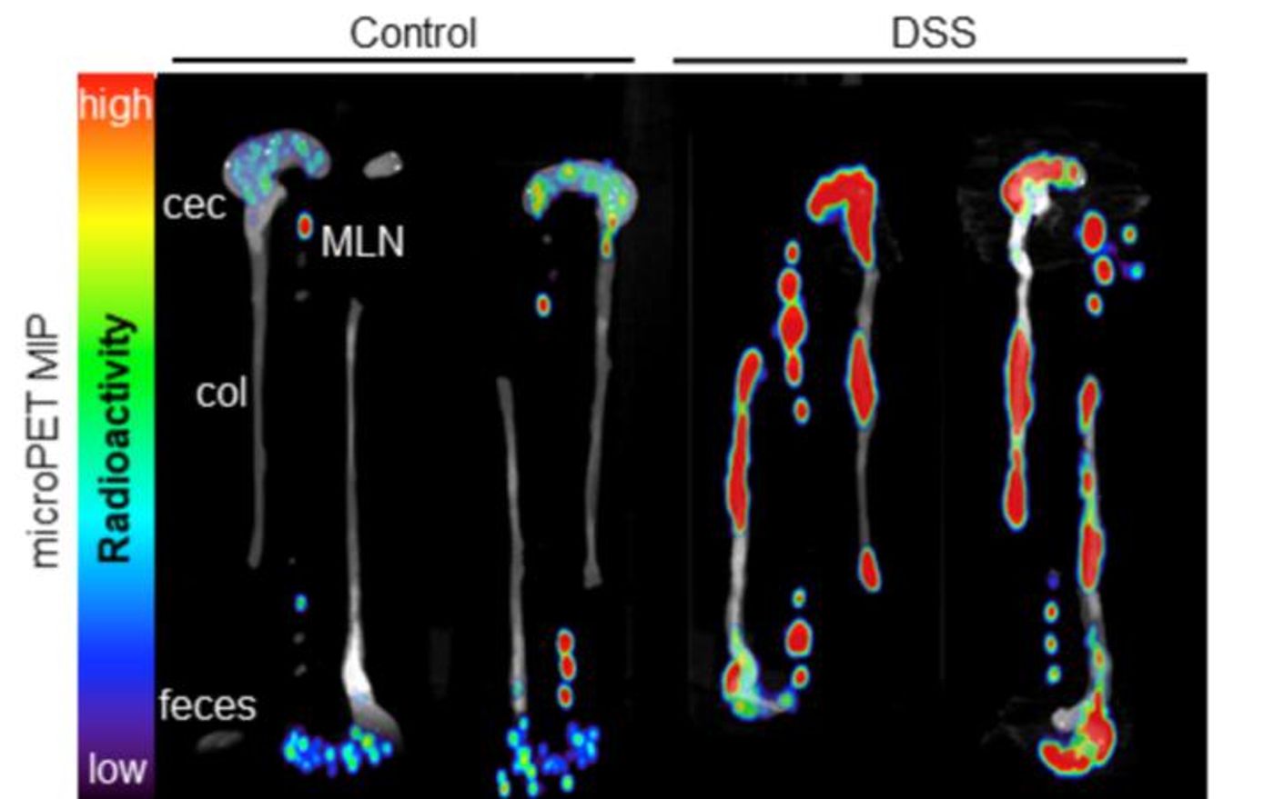
Wu and others tested immunoPET with a mouse model of induced colitis, observing the distribution of CD4+ T cells. The imaging results revealed T cells in the colon, ceca, and mesenteric lymph nodes. The ceca is the part of the GI tract at the beginning of the large intestine.
Going forward, the study researchers see immunoPET as an opportunity to create specialized antibody-based imaging for human IBD patients.
"It could unlock our ability to assess inflammation in a broad spectrum of disease areas, including oncology and immune-oncology, auto-immunity, cardiovascular disease, neuroinflammation, and more,” Wu explained. “ImmunoPET is a robust and general platform for visualization of highly specific molecular targets."
The present study was published in the Journal of Nuclear Medicine.
Sources: Blood, RadiologyInfo.org, Society of Nuclear Medicine and Molecular Imaging