A CT scan can do more than just reveal broken bones. In a new study, researchers found that CT scans can provide a glimpse into a patient’s overall muscle health, as well as predict survival following hip fractures.
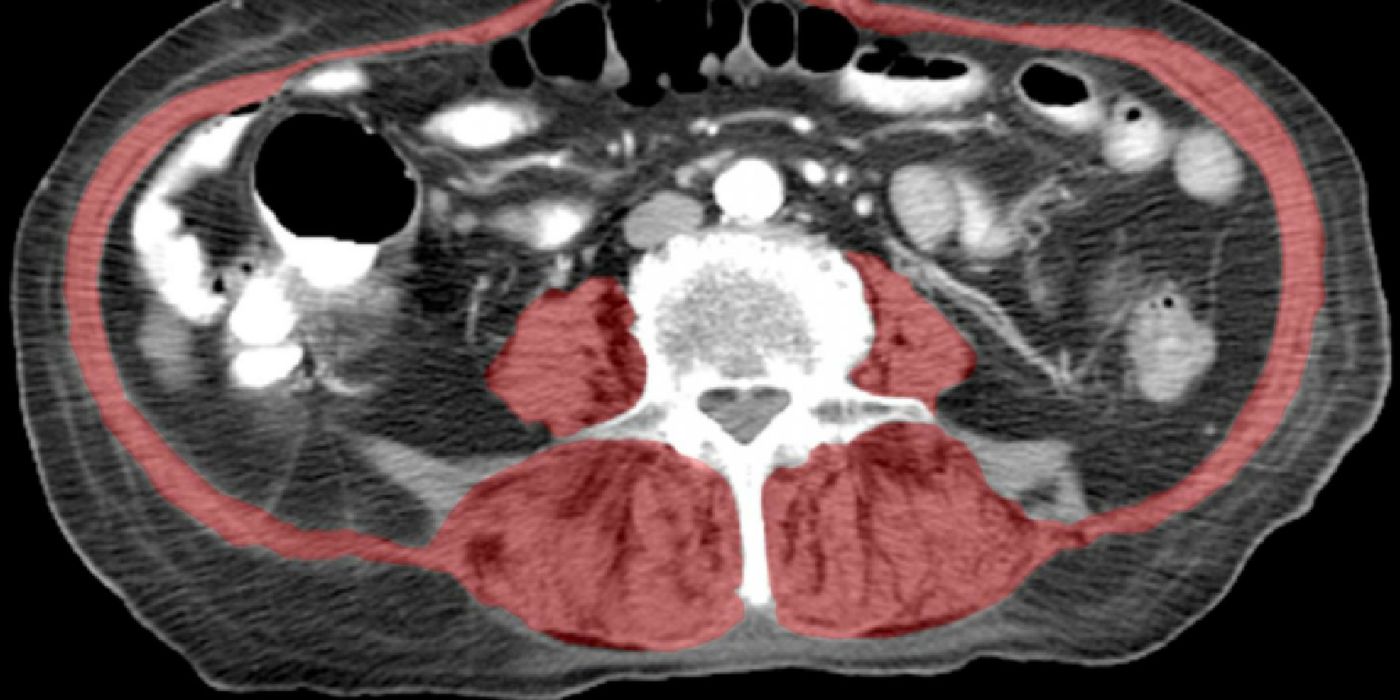
Computed tomography (CT) scans combine a series of X-ray images taken at multiple angles to create a cross-sectional image of the body. This technology is widely used to diagnose bone and muscle conditions, and to detect bleeding and cancer.
But more than broken bones, the CT images reveal the health of the surrounding muscles. In particular, the scans give away the density of the “core” muscles that stabilize the spine. Those with lower core muscle size and density are more likely to be more frail, and therefore, have worse outcomes than patients whose core muscles are in better shape.
This knowledge is tremendously important when talking about the elderly population. "As patients age, it becomes increasingly important to identify the safest and most beneficial orthopaedic treatments, but there currently is no objective way to do this," said Robert Boutin, a UC Davis professor of radiology, and the study’s lead author.
Together with his colleagues, Boutin studied 300 elderly patients, aged 65 and older, who were treated for fall-related injuries between 2005 and 2015 at the UC Davis Medical Center. These patients all received CT scans to confirm or rule out hip fractures. Boutin then compared the images with mortality data from the National Death Index.
They found that not only can muscle information from CT scans predict the outcome of the treatment, the information also predicted survival. People who showed better core muscles had better survival rates than those with poor core muscles.
"Using CT scans to evaluate muscles in addition to hip bones can help predict longevity and personalize treatment to a patient's needs. We're excited because information on muscle is included on every routine CT scan of the chest, abdomen and pelvis, so the additional evaluations can be done without the costs of additional tests, equipment or software,” said Boutin.
This is the first study to link CT scans of hip fractures with survival in a non-cancer population. "The fact that we were able to predict survival in such a small group of non-cancer patients is truly remarkable," said Lenchik, professor of radiology at Wake Forest, and the study’s senior author.
"Recognizing sarcopenia [muscle loss] as a distinct condition that provides clues to future health can open doors to new discoveries in diagnosis and treatment," Boutin said. The team hopes such results will be leveraged to provide better treatment options for patients.
Additional source: UC Davis Medical Center