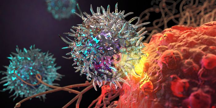
Currently, if a doctor suspects a patient has cancer or other diseases, he orders a tissue biopsy to confirm whether the pathology of the sample matches the diagnosis. The biopsy can be highly invasive, costly, and require weeks to months for processing. To circumvent this routine, researchers at the Imperial College London have engineered two microscope prototypes that can assess the tissue and take images in the patient in real time. The team hopes their instruments can reduce stress to the patients, while simultaneously cutting time and healthcare costs.
Of the two devices, the first is a handheld microscope that doctors can use to assess external tissues. This device can even be used by surgeons during surgery, letting doctors know, in real time, whether the exposed tissues is cancer or normal. Key to this microscopes innovation is its internal tracking mechanism that makes it possible to acquire high quality 3D images of tissues. This means that tissues can be analyzed within a breathing, moving patient, instead of pre-extracted tissues.
The second prototype is an endoscope – a lit device that lets doctors see inside cavities of the body without drastic surgery. Coming at a fraction of a millimeter in diameter, the freshly designed endoscope uses optical fibers and can be threaded through tiny areas with minimal effort. It will allow doctors to see at the cell level even deep inside the body.
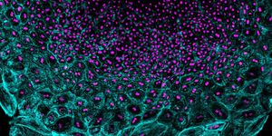
Both devices rely on a technique known as multiphoton excited fluorescence microscopy, which involves “involves illuminating tissue with light of a specific wavelength that causes molecules in the tissue to emit light in response,” according to the Engineering and Physical Sciences Research Council release. As such, the devices allow researchers to obtain high resolution images at impressive depths beneath the surface of the body, all while the patient is on the table.
In addition to cutting down the time it would take to process samples and confirm a diagnosis, the data obtained by the prototypes could, arguably, be considered more reliable. This is because doctors can view the cells in real time, in the body’s native context. Thus, their conclusions on whether a tissue is abnormal are free from the effects of specimen processing.
"These new devices open up exciting possibilities in the field of in-situ diagnosis and could help improve patient care in the future," said Chris Dunsby, researcher at the Imperial College London, and study leader.
"This has been a very exciting project which has enabled us to develop fibers with performance which we would have previously thought impossible," said Jonathan Knight, study leader from the University of Bath.
Additional source: Engineering and Physical Sciences Research Council