Move over, mammograms. University of Michigan scientists have a new solution for diagnosing breast cancer that’s possibly more accurate, less invasive, and less uncomfortable. With a pill that delivers a dye to illuminate tumors under infrared light, determining the difference between benign and malignant could become easier than ever before.
Many women undergo unnecessary diagnostic tests only to find that their tumor is harmless. The new technology might improve the chances that invasive approaches like surgery and chemotherapy can be reserved for malignant tumors that require this step. Additionally, scientists want to limit the malignant tumors that go unnoticed because thick breast tissue hides lumps. And of course, limiting the number of unnecessary tests will help avoid unnecessary costs.
"We overspend $4 billion per year on the diagnosis and treatment of cancers that women would never die from," explained study leader Greg Thurber, PhD.
The new diagnostic method provides an alternative to mammography, which is uncomfortable for many women and which experts from the study say is imprecise. Mammography provides an x-ray image of the breast, obtained by flattening the breast against the mammogram machine.
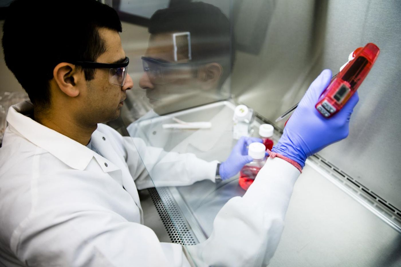
University of Michigan researchers, led by Thurber, developed a pill, taken orally by the patient being tested, that delivers a dye responding to infrared light. The dye tags a molecule commonly found on tumor cells and in the surrounding environment. The information obtained from this test can answer the most important question: benign or malignant?
While developing the new diagnostic system, researchers came across issues with creating a pill to carry the dye to the tumor.
"To get a molecule absorbed into the bloodstream, it needs to be small and greasy,” Thurber explained. “But an imaging agent needs to be larger and water-soluble. So you need exact opposite properties.”
They found their solution in a targeting molecule originally designed by Merck for treating cancer. While the molecule didn’t work for their purposes, it did work for the University of Michigan scientists. With this targeting molecule incorporated, the pill can successfully travel through the stomach to the liver and into the bloodstream.
For now, researchers have only shown the diagnostic method to be effective and safe in mice, but further studies are definitely on the horizon.
The present study was published in the journal Molecular Pharmaceutics.
Sources: American Cancer Society, University of Michigan