In 2016 a relatively simple burial of two individuals was excavated at the site of Tel Megiddo. I say relatively simple because immediately adjacent to it was another tomb containing seventeen people with gold, silver, bronze, and more. In this case, the simpler Tomb 45 included two adult males determined to be brothers through genetic analysis of the skeletal remains.
Unlike the more elaborate burial next door, these brothers were buried with food and ceramics, not gold and silver. However, this burial is still considered high-status due to the burial context.
The brothers were not buried at the same time. Archaeological evidence suggests that Individual 2 was buried first, then exhumed and re-buried when Individual 1 was interred. This type of grave disturbance and re-inhumation is not uncommon across variable archaeological contexts.
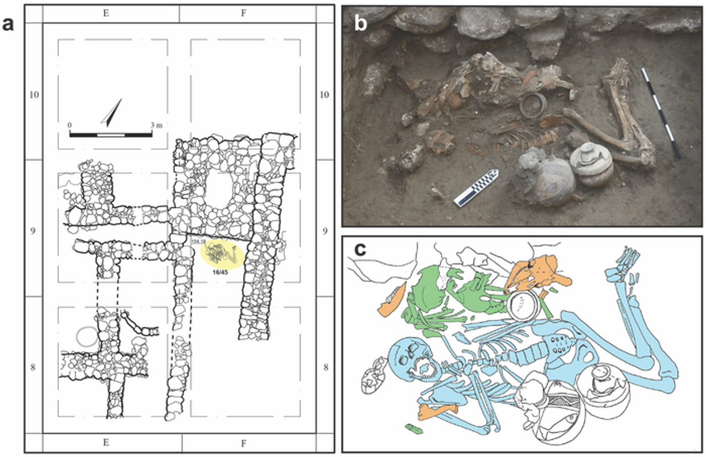
Image showing: a. Tomb 45 location highlighted in yellow; b. photograph of the burial; c. composite drawing of the burial with Individual 1 in blue and Individual 2 in Green. Credit: Kalisher et al., 2023, PLOS ONE, CC-BY 4.0 (https://creativecommons.org/licenses/by/4.0/).
The skeletal remains of both Individual 1 and Individual 2 share not only signs of inherited congenital conditions but also show signs of widespread infectious disease. Paleopathologically speaking, long-term infectious diseases can leave a mark on human bone. Some, like Venereal Syphilis and Leprosy, have distinctive features specifically suggestive of those diseases. For example, caries sicca, a star-like lesion found on the crania, is diagnostic of venereal syphilis infections. However, unfortunately for paleopathologists and bioarchaeologists everywhere, you can’t always count on finding these diagnostic features in every case of syphilis in archaeological populations. Variability in disease progression and skeletal preservation make concrete diagnosis difficult in many cases.
In this case, both individuals displayed various types of bone lesions spread across the cranial and postcranial skeleton. Unfortunately, damage to the remains precludes any exact diagnosis, but the authors offer potential differential diagnosis options, including osteomyelitis, treponemal disease (like venereal syphilis), tuberculosis, and leprosy. While it’s not possible to be exact here, it’s evident that both individuals suffered from a long-term, chronic, infectious disease.
The identification of the congenital and infectious conditions in these siblings is fascinating, but even more interesting (at least in my opinion) is the potential ‘treatment’ of disease in Individual 1.
The skull of Individual 1 had a square piece of bone that was removed from the frontal bone immediately prior to death. This procedure, trephination, is the removal of bone from the skull by one of several methods. In this case, the bone was notched and cut away in pieces which were also found within the burial. This procedure has been identified across the ancient world to varying levels of success and was performed to treat traumatic injuries, diseases, and as a part of ritual practices.
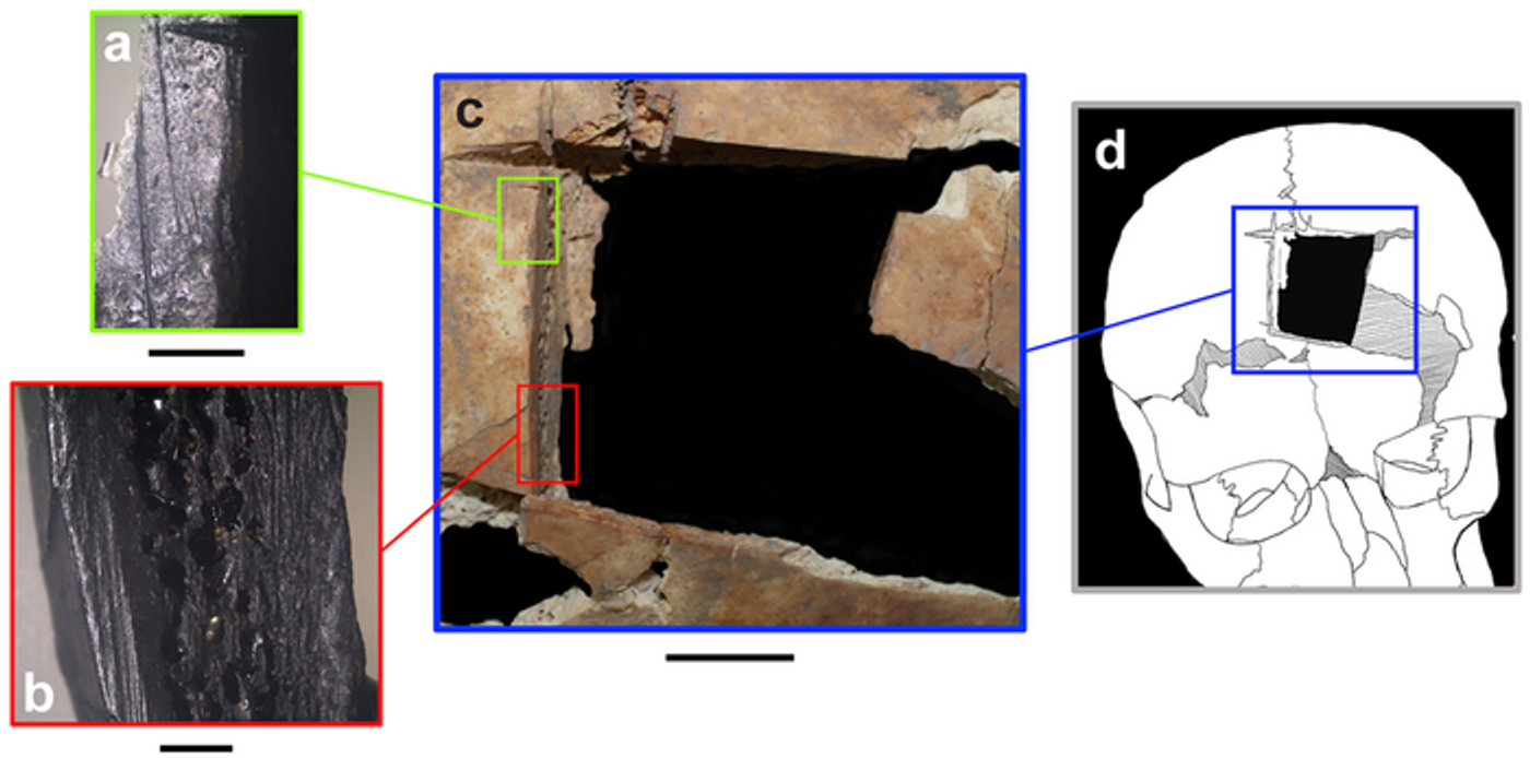
Image showing: the trephination of Individual 1 at the macroscopic level (left) and in composite (right). Credit: Kalisher et al., 2023, PLOS ONE, CC-BY 4.0 (https://creativecommons.org/licenses/by/4.0/).
The authors note that “Among the study’s multiple findings, we wish to highlight the special type of cranial trephination, the earliest of its kind in the region. This uncommon procedure was done on an elite individual with both developmental anomalies and infectious disease, which leads us to posit that this operation may have been an intervention to deteriorating health.”
Unfortunately, in this case, the procedure was unsuccessful. Bone healing was not observed at the microscopic level (1-2 weeks post-op) or macroscopic level (2-5 months post-op), suggesting that the individual died during or soon after the procedure.
These individuals stand out in their health and social status. However, their burial was not unique; in fact, it was very similar to others of the same time period. They were not “othered” by their conditions or, in the case of Individual 1, the potential manner of their death, an exciting feature that may help us better understand the treatment of the sick at different levels of society.
Sources: PLoS One, Science Direct: Paleopathology, Johns Hopkins Medicine, Science Direct: Trephination, MIT Press, EurekAlert!
Open Access Paper Available in PLoS One: Kalisher R, Cradic MS, Adams MJ, Martin MAS, Finkelstein I (2023) Cranial trephination and infectious disease in the Eastern Mediterranean: The evidence from two elite brothers from Late Bronze Megiddo, Israel. PLoS ONE 18(2): e0281020.
Featured/Cover Image Credit: Credit: Kalisher et al., 2023, PLOS ONE, CC-BY 4.0 (https://creativecommons.org/licenses/by/4.0/).