Scientists at the Université de Montreal (UdeM) have used super-resolution microscopy to get a new look at the three-dimensional structure of messenger RNA molecules. Their research upends how we think of these critical molecules, which transmit information from the genome to cellular machinery, which uses it to make proteins. The study, which has been reported in Molecular Cell, shows that the conformation of mRNA changes according to where in the cell it's located.
The video illustrates how DNA is made into proteins, and the role mRNA plays in the process.
"The flow of information from DNA to protein implicates a copy of the DNA sequence called messenger RNA that serves as template for protein synthesis," said the study's senior author Daniel Zenklusen, an associate professor at UdeM's department of biochemistry and molecular medicine. "Just like DNA, RNA is a long polymer composed of nucleic acids. How these RNA polymers are compacted and organized in cells to allow protein synthesis was so far unknown, in part because we were lacking technologies to visualize these molecules in high resolution," Zenklusen said.
It had been thought that when the cell is using mRNA to make proteins, the two ends of the mRNA molecule come together to form a specific structure, called a closed-loop complex. This work has found that to be an oversimplification.
"We observed that messenger RNAs exist in many different configurations in cells, but not in the previously suggested stable closed-loop conformation," said the first author of the study, Srivathsan Adivarahan, a graduate student in Zenklusen's lab. "This was very surprising to us since this model is found in every textbook describing the essential process of protein synthesis."
The researchers worked with teams led by Olivia Rissland at the University of Colorado and Bin Wu at Johns Hopkins University and learned that mRNA exists in a variety of forms, but for the most part the molecule remains very compact. The mRNAs are at their smallest when protein synthesis has been halted, or when they are relegated to specific parts of the cell.
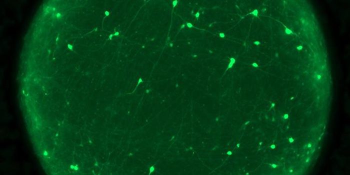
In the video from Johns Hopkins University (not related to this work), messenger RNA, seen in green, builds up in one part of a neuron as translation is suppressed.
"Our findings change how we think about many aspects of mRNA metabolism, and in particular on how the mRNA is organized during protein synthesis," said Zenklusen. Regulating this process is essential for all cells, but it is particularly important for a cancer cell that requires high levels of protein synthesis to allow for unceasing growth. Therefore, different drugs affecting proteins synthesis are currently in development and some of these drugs target proteins previously implicated in the closed-loop model. The models of how these drugs affect protein synthesis will have to be revisited."
This work illustrates that basic science studies are still an important part of innovation, he added. "Technological advances allow us to revisit questions we long thought to have been solved, just to realize once we look at them with new eyes that we are far from truly understanding them."
The researchers are planning to continue this work. The Zenklusen laboratory wants to improve microscopy techniques used at the molecular level to gain more insight into how gene expression is controlled, and how it goes wrong in disease.
Sources: AAAS/Eurekalert! via UdeM, Molecular Cell