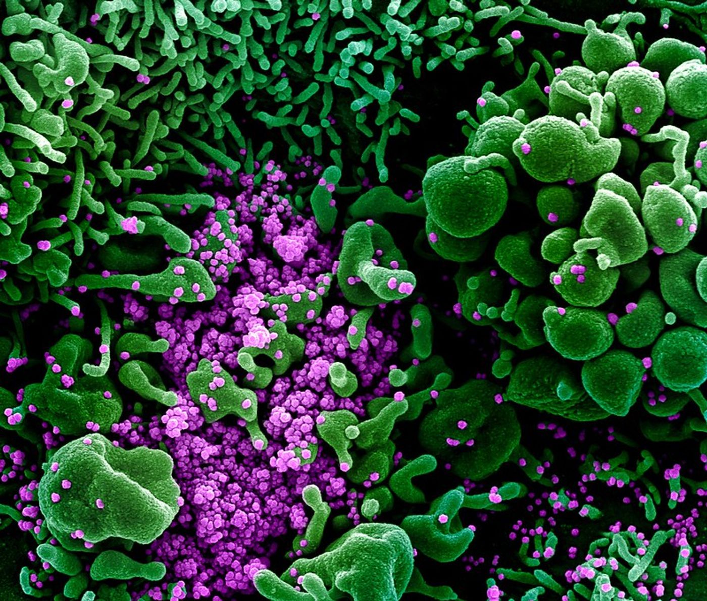
PCR (polymerase chain reaction) is a fundamental tool used in many aspects of biological research. In the reaction, a specific piece of DNA can be identified and amplified using short strands of nucleotides called primers. These primers can amplify DNA of interest from very small quantities of template DNA in a sample. For example, a patient's blood can be tested for COVID-19 by using primers that are specific to the genetic material of SARS-CoV-2. If the virus is present, the primers will find it and will amplify it so it can be detected.
Identifying people who have the virus and testing their contacts will be the fastest way to get society functioning again. Unfortunately, however, huge numbers of these PCR-based tests would be needed to do that, and this massive demand may overwhelm the supply of PCR reagents. These tests can also be unreliable; there have been reports of false-positive and false-negative results.
To ease the strain on PCR reagents and the machines that are used to carry out the reactions, scientists have been looking for other ways to diagnose the virus, including CT scans and antibody tests. Researchers have now created a diagnostic test based on a concept called plasmonic photothermal sensing. The work has been reported in ACS Nano.
In this method (explained in the video), a beam of light hits a metallic surface, and that beam then bounces onto a detector. The metallic surface is a sensor chip, and the electrons inside get excited by the light beam hitting the surface; they resonate. In this case, the sensor chip holds probes that recognize sequences of SARS-CoV-2 RNA. These probes are linked to gold nanoparticles. When pieces of the viral genome are present in a sample, the viral RNA will link up with the probes, and by applying heat, false-positives are reduced.
When the researchers tested their method, the assay was very specific; it was able to tell the difference between SARS-CoV-2 and a close viral relative, SARS-CoV-1. One drawback of the diagnostic test, however, is that it requires intact viral RNA from patient samples.
Experienced research scientist and technical expert with authorships on over 30 peer-reviewed publications, traveler to over 70 countries, published photographer and internationally-exhibited painter, volunteer trained in disaster-response, CPR and DV counseling.