Genetics & Genomics
Neanderthal DNA Helps us Fight Viruses
OCT 08, 2018 7:52 AM PDT
Share
Nuclear Imaging: The Next Generation of Biopsy
Organ transplantation is a significant procedure that brings the potential for complication. Often, our immune systems can overact to the new organ and reject the transplantation. The immune system will recognize the organ as a foreign substance that it must eliminate, resulting in a lifetime of rigorous treatment to stave off the negative impacts of the procedure. For reasons such as these, physicians seek to get ahead of the risk by closely monitoring the organ function.
A team of researchers has published their work in the Journal of Nuclear Medicine describing techniques are ultimately be safer for patients. Usually, to determine the functionality of an organ or tissue, physicians will make use of biopsy methods. However, biopsy procedures are invasive and not without potential for complication. Positron-emission tomography or PET imaging methods were explored as an alternative to a biopsy.
PET imaging is described as a scan that will reveal how tissues and organs are functioning. The PET imaging makes use of a radioactive drug (the tracer) to highlight activity. PET imaging can detect disease before it appears on other imaging tests.
Depending on the tissue or organ being observed, tracers are injected, swallowed or inhaled. The tracer will collect in areas of the body that have high levels of chemical activity, which often correspond to regions of disease. The imaging will display on a monitor as bright spots.
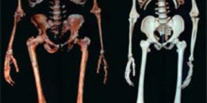
In the study performed by researchers at the University of California and University of Pennsylvania, they utilized a molecule called 18 F-FAC as their radioactive tracer to observe liver function. The creation of a mouse model allowed for testing of T cells in the liver. T cells in the liver are looked for in this case because of their role clearing foreign substances from the body. Thus, their presence would indicate the potential for rejection of a transplanted liver.
"The personalized treatment of patients suffering from immune attack on the liver will require precise and quantitative methods for measuring T cells in the liver," said Peter Clark, Ph.D. "Our work describes in a preclinical model one such way of doing this. We envision that if this approach is translated into the clinic, it could lead to fewer liver biopsies and more precise treatment of patients with immune-related liver disease."
The research of molecular imaging performed here might have the potential to be applied in diagnoses of other chronic liver infections.
The data from this study suggest that PET can be used to noninvasively visualize activated T cells and inflamed liver cells in mice with autoimmune hepatitis.
Sources: Journal of Nuclear Medicine, YouTube, Mayo Clinic

Master's (MA/MS/Other)
You May Also Like
OCT 06, 2025
Cancer
Lymph nodes, hundreds of small structures located throughout the body, play a pivotal role in immunity, including anti-t
...
NOV 20, 2025
Immunology
Immunotherapy has revolutionized the way physicians treat patients. Previously, chemotherapy was the standard-of-care wi
...
NOV 26, 2025
Genetics & Genomics
Mitochondria are well known as the powerhouses of the cell because of their energy generating capabilities. These little
...
JAN 15, 2026
Cannabis Sciences

How can cannabidiol (CBD) combat cancer tumors? This is what a recent study published in Phytomedicine hopes to address
...
JAN 26, 2026
Cell & Molecular Biology

The immune system is a powerful, necessary shield that has to spring into action when an infection has to be eliminated.
...
FEB 09, 2026
Cancer

Research has suggested that cancer treatments administered early in the day are more effective than those administered l
...
Loading Comments...