Microglia hold down the fort in the brain as the primary immune cell presence. When brain disease hits, they get rid of injured neurons by breaking their synapses, their connection to other neurons, in order to protect the brain against disease. Even though microglia make up 10 percent of the brain's cells, until 2005 scientists thought that the microglia only came out to play when a foreign invader threatened the brain's safety.
After research studies showed microglia to be the fastest moving cells in a
healthy adult brain, other studies began to figure out just what extra role the microglia were fulfilling. More studies produced results showing interactions between microglia and neuron synapses in healthy brains.
Similarly to peripheral macrophages, microglia produce cytokines and chemokines to induce the immune response during brain injury (
University of Carlifornia, Irvine). They are also the cells that manage the immune response once a pathogen has been successfully contained, secreting interleukin 10 and other anti-inflammatory cytokines to stop inflammation.
Neuroscientists at a lab in
Boston's Children's Hospital looked into microglia action during early brain development. Microglia were found to be eliminating unnecessary neuron synapses, since the brain initially develops with more than it needs. Dr. Emily Lehrman discovered a "protective tag" that communicates to microglia which synapses to leave alone so the immune cells don't eliminate too many - similar to how immune cells recognize host cells as "self" when patrolling the body looking for foreign invaders.
"Excess engulfment by microglia and overpruning of neuronal connections" occurred when experimental mice had their protective tag removed.
Another group at the
European Molecular Biology Laboratory in Germany is looking at the role of fractalkine, a molecule heavily involved in neuron-microglia signaling. Fractalkine receptors are found only on microglia, and experimentally removing these receptors cause the microglia to neglect deleting weak synaptic connections like they normally do in early development. Microglia also mature similarly to neurons, further insinuating a connection between neurons, fractalkine, and microglia.
In this studies when microglia did not delete weak synaptic connections, brain development was hindered and signs of autism occurred. Scientists are not yet certain whether this connection is a cause or effect relationship.
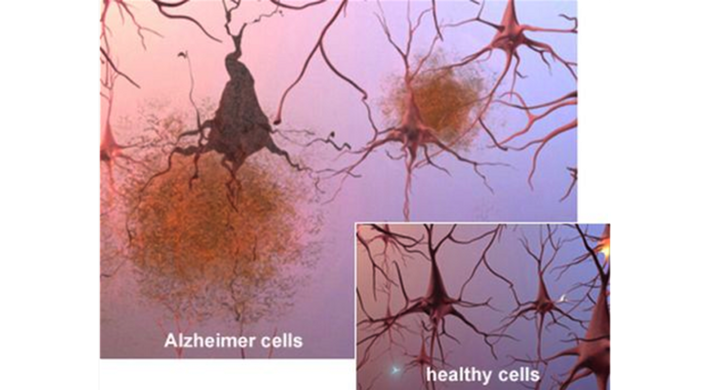
As part of their role of deleting dead and dying cells during disease, microglia have also been found to be ingesting and disposing of beta-amyloid deposits in cases of Alzheimer's disease. This discovery could be the microglia delaying
or exacerbating the progression of Alzheimer's.
A lot is still unknown about the precise role of microglia as the immune cells of the brain. There is a connection, though, and as soon as scientists can determine how to manipulate this mechanism, there will be a lot of areas in which to create new therapeutics.
Watch the following video to see "microglia in action" amongst neurons in the brain.
Source:
Scientific American