
Clinical & Molecular DX
Software Flags Elevated Cerebral Palsy Risk in Premies
Interested in health technology and innovation.
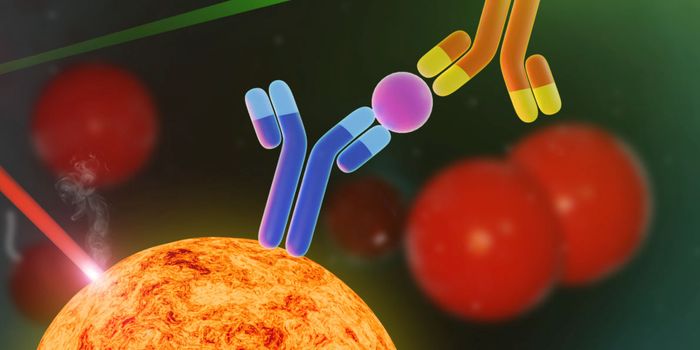
A team of researchers at Duke University have developed an imaging technology for tagging structures at a cellular level that overcomes the shortcomings of existing antibody-based techniques. Immunofluorescence imaging is a key part of the cell biologist’s toolbox, in which a fluorescent ‘flare’ attached to an antibody allows them to visualize the presence of specific target proteins in cell or tissue samples. The issue is that this specificity isn’t always 100 percent — sometimes the antibodies bind to other closely related proteins as well, making it difficult to interpret the results.
Duke’s cell biology chair Scott Soderling has led a team that developed Homology-independent Universal Genome Engineering (HiUGE), an innovation that uses gene-editing technology to rise above the shortcomings of traditional commercial antibodies for imaging.
“We had this idea that CRISPR could be a really amazing tool to address the pressing problem of trying to identify and label these hundreds of proteins,” said Soderling.
“What we developed was a new modular method for basically taking the labeling problem and flipping it on its head.”
HiUGE uses CRISPR to insert a short genetic sequence into the target protein’s gene using an adenovirus delivery system. This molecular tag which codes for a segment of amino acid sequences is detected by a highly sensitive antibody which is significantly more reliable than many of the commercially-available antibodies for research on the market. Different combinations of these CRISPR tags have the potential to mark hundreds of proteins and the technology can easily be adapted to automated, high-throughput laboratory imaging processes.
Neuronal signaling has been a key focus of Soderling’s research and the discovery of HiUGE is allowing the team to “see” these intricate processes in the brain like never before. Relying on antibodies has been a barrier, in part because they fail to visualize the complex myriad of molecular events occurring at the synaptic junction at high enough resolution. HiUGE can be administered to mice, allowing scientists to map and track the dynamics and localization of neural proteins with stunning clarity as depicted in the video.
Sources: Neuroresource, Duke University.