Optogenetics is a technology used by neuroscientists to follow and record the course of individual neurons as they fire. Using different colors of light to stimulate activity, scientists can learn more about the inner workings of animal brains. Scientists from the University of Massachusetts Medical School, in collaboration with scientists from Texas A&M Science Center Institute of Biosciences & Technology, thought that this technology could also be incorporated into a new cancer immunotherapy, and they were right.
Although lymphocytes do not fire action potentials to communicate with each other like neurons do, they have their own methods for cell signaling. Together, scientists from both universities developed an optogenetic immunomodulation technology that relies on the regular flow of calcium ions through channels on the surface of dendritic cells.
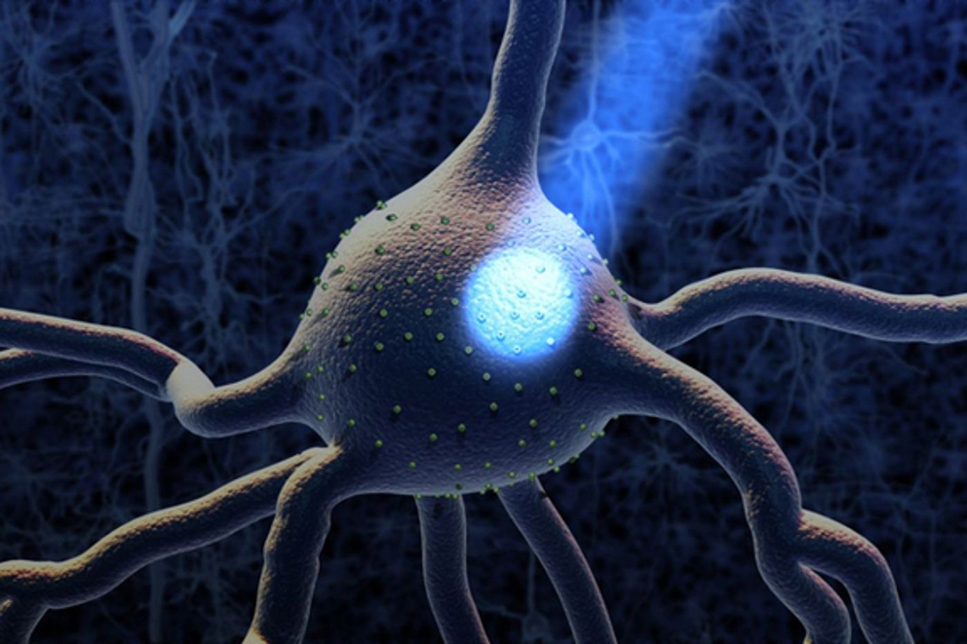
Dendritic cells play a vital role in an immune attack. By responding to infection with displaying antigens on their cell surface, T cells are alerted of an invasion and instruct B cells to make antibodies targeting the invader. Scientists conducting this study, published recently in the journal
eLife, used mouse models of melanoma to see if their new optogenetic immunomodulation technology could activate dendritic cells to attack melanoma tumors.
First, Texas A&M scientists equipped dendritic cells with a light-sensitive calcium gate-controlling protein. These cells’ calcium channel gates then respond to blue light, either opening with the light on or closing with the light off. When the gates open, calcium ions flood in and activate the dendritic cell. With the light off, the gates close and the dendritic cell returns or stays in a dormant-like state.
Next, University of Massachusetts scientists made these equipped dendritic cells accessible to light within an animal by developing a nanoparticle to attach to the cells that converts “near-infrared light” into the visible blue light to be used for controlling calcium ion channels. The near-infrared light can infiltrate tissue up to two centimeters. The dendritic cells and attached nanoparticles are called “opto-CRAC.”
“We now have the tools to closely monitor the dose and location of [cancer] treatment to mitigate potential side effects to healthy tissues,” said author of the study Gang Han, PhD, from the University of Massachusetts.
With their technology in place, the team was able to test their control over dendritic cell activation with calcium ion channel regulation. They introduced opto-CRAC into mouse models of melanoma as well as the “tumor antigen surrogate” ovalbumin. The tumor antigen component is required for dendritic cell activation of T cells, should the new technology prove to be successful.
The new optogenetic immunomodulation technology was indeed successful:
"We saw significantly suppressed tumor growth and reduced tumor volume in these animals,” Han said. “This suggests that the activated dendritic cells were successfully programming T-cells to attack the tumor.”
Further success with this technology could lead to less invasive cancer treatments, since specific cells and tissues compromised by cancer can be targeted, and healthy tissue can be left alone.
Source:
University of Massachusetts Medical School